Multicenter Study
. 2022 Jul;63(4):393-402.
doi: 10.1111/vru.13063. Epub 2022 Jan 20.

Imaging features of retroperitoneal extra-adrenal paragangliomas in 10 dogs

Affiliations
Multicenter Study

Imaging features of retroperitoneal extra-adrenal paragangliomas in 10 dogs

Alexis Gombert et al. Vet Radiol Ultrasound. 2022 Jul.

Abstract

Retroperitoneal paragangliomas are rare tumors of the neuroendocrine system. Only a few canine case reports are available with rare descriptions of their imaging features. The objectives of this multi-center, retrospective case series study were to describe the diagnostic imaging features of confirmed retroperitoneal paragangliomas and specify their location. Medical records and imaging studies of 10 affected dogs with cytological or histopathologic results concordant with retroperitoneal paragangliomas were evaluated. Dogs had a median age of 9 years. Four of them had clinical signs and laboratory reports compatible with excessive production of catecholamines. Six ultrasound, four CT, four radiographic, and one MRI studies were included. The paragangliomas did not have a specific location along the aorta. They were of various sizes (median 33 mm, range: 9-85 mm of length). Masses had heterogeneous parenchyma in six of 10 dogs, regardless of the imaging modality. Strong contrast enhancement was found in all CT studies. Encircling of at least one vessel was detected in six of 10 masses, clear invasion of a vessel was identified in one of 10 masses. In five of 10 cases, the masses were initially misconstrued as lymph nodes by the on-site radiologist. Retroperitoneal paragangliomas appear along the abdominal aorta, often presenting heterogeneous parenchyma, possibly affecting the local vasculature, and displaying strong contrast enhancement on CT. Clinical signs can be secondary to mass effects or excessive catecholamine production. Underdiagnosis and misdiagnosis of this tumor are suspected as they can be silent, of small size, or confused with other structures.

Keywords: MRI; canine; computed tomography; neuroendocrine; ultrasound.


Conflict of interest statement

The authors have declared no conflict of interest.

Figures
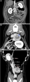
FIGURE 1
FIGURE 1
Computed tomography appearance of different retroperitoneal paragangliomas. Left is to the right. A, Case 4. Dorsal multiplanar reconstruction in minimal intensity projection in arterial phase after intravenous contrast injection in a 7‐year‐old neutered female Shih‐Tzu displaying an oval mass with strong heterogeneous contrast‐enhancement, multifocal mineralization foci, and close contact (grade‐4) with the right renal vein (arrow), and ipsilateral adrenal gland (+). B, Case 6. Post‐contrast CT images of a retroperitoneal paraganglioma in an 8‐year‐old German Shepherd Dog. Transverse plane in venous phase after intravenous contrast injection showing a rounded heterogeneous mass with mineralization focus (*) in close contact with three vessels (arrows): from dorsal to ventral, grade‐2 with the aorta and caudal vena cava and grade‐4 with the left renal vein. Example of ROI placement is demonstrated by the blue circle. C, Dorsal postcontrast multiplanar reformatted CT image illustrating the topography of the tumor (arrowhead) located caudally to the left renal artery (Renal a.). Note the caudal position and distance compared to the ipsilateral adrenal gland (Left AG) located caudally to the celiac (Celiac a.) and cranial mesenteric arteries (Cr. Mes. A.). The contact with the caudal vena cava (*) is visible. Images reconstructed with standard algorithm and displayed with a window width and window level of 40 HU and 400 HU. Images acquired in sternal recumbency, with a kVp of 120 (A,B,C), mA of 412 (A), 319 (B,C), and a slice thickness of 0.9 mm (A), 2.5 mm (B,C) [Colour figure can be viewed at wileyonlinelibrary.com]
FIGURE 2
FIGURE 2
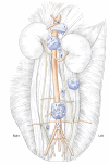
Representations of the different retroperitoneal masses of all included dogs. Each mass number matches the case number as shown in the Supporting Information 1 [Colour figure can be viewed at wileyonlinelibrary.com]
FIGURE 3
FIGURE 3
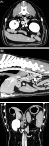
Postcontrast CT images of a retroperitoneal paraganglioma in case 7, a 13‐year‐old crossbreed neutered male dog. A, Transverse post‐contrast CT image illustrating the tumor (arrowhead) demonstrating homogeneous contrast enhancement. Note the close contact with the caudal vena cava (*) and aorta (#) (both grade‐2). B, Sagittal postcontrast multiplanar reformatted CT image illustrating topography of the tumor (arrowhead) located medially to the left adrenal gland (AG), ventrally to the aorta (#), caudally to the cranial mesenteric artery. C, Dorsal postcontrast CT image illustrating normal appearance of the left adrenal gland (Left AG) and medial position of the paraganglioma (arrowhead), ventrally to both aorta (#) and caudal vena cava (*). Images reconstructed with standard algorithm and displayed with a window width and window level of 40 HU and 400 HU. Images acquired in sternal recumbency, with a kVp of 120, mA of 319 and a slice thickness of 2 mm
FIGURE 4
FIGURE 4
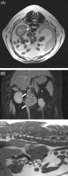
MRI acquisition (1.5 T) of the case 3, a 7‐year‐old neutered female Boxer dog. A, Transverse T2‐weighted Spin‐echo sequence, (TR: 5670 ms, TE 109 ms), 6‐mm slice thickness. The mass is amorphous with ill‐defined margination, heterogeneous and hyperintense compared to paraspinal muscles. It is here slightly right‐sided. The caudal vena cava (arrow) is severely compressed. The right adrenal gland is dorsal (arrowhead) within normal limits. B, Dorsal T1‐weighted with fat saturation sequence after intravenous contrast medium injection, (TR: 935 ms, TE: 14 ms) 6‐mm slice thickness. The mass is heterogeneous and compresses the caudal vena cava (arrows): grade‐6. C, Sagittal T1‐weighted Spin‐echo sequence, (TR: 246 ms, TE: 14 ms), 3‐mm slice thickness: the mass is hypointense compared to paraspinal muscles and demonstrates irregular margins
FIGURE 5
FIGURE 5
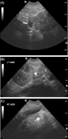
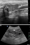
Ultrasonographic appearance of the mass of Case 9, (microconvex probe, 11 MHz) a 9‐year‐old neutered male Boxer dog: A, Lobulated hypoechoic mass with invasion of the caudal vena cava (arrow) is identified. B, The mass (‡) is in close proximity to the left adrenal gland (LT ADR, arrowhead), within normal limits. C, The contralateral adrenal gland (arrowhead) is within normal limits (RT ADR: right adrenal gland)
FIGURE 6
FIGURE 6
Ultrasonographic appearance of different retroperitoneal paragangliomas. A, Case 4, a 7‐year‐old neutered female Shih‐Tzu (linear probe, 18 MHz). Oval heterogeneous nodule showing mineralization (*) and close contact with the caudal vena cava (arrow). B, Case 8. Microconvex probe, 8 MHz. Small rounded homogeneously hypoechoic nodule (arrowhead) representing a paraganglioma suspected to be hormonally active in 13‐year‐old crossbreed neutered male dog. The left renal artery is on the right of the image and left phrenicoabdominal artery on the left [Colour figure can be viewed at wileyonlinelibrary.com]

References

    1. Gill T, Adler K, Schrader A, et al. Extra‐adrenal pheochromocytoma at the organ of Zuckerkandl: a case report and literature review. Radiol Case Rep. 2017; 12:343‐347. - PMC - PubMed
    1. Moonim MT. Tumours of chromaffin cell origin: phaeochromocytoma and paraganglioma. Diagn Histopathol. 2012; 18:234‐244.
    1. Lam AK. Update on adrenal tumours in 2017 World Health Organization (WHO) of endocrine tumours. Endocr Pathol. 2017; 28:213‐227. - PubMed
    1. Galac S, Korpershoek E. Pheochromocytomas and paragangliomas in humans and dogs. Vet Comp Oncol. 2017; 15:1158‐1170. - PubMed
    1. Robat C, Houseright R, Murphey J, et al. Paraganglioma, pituitary adenoma, and osteosarcoma in a dog. Vet Clin Pathol. 2016; 45:484‐489. - PubMed

Publication types

MeSH terms