. 2022 Feb 18;375(6582):760-764.
doi: 10.1126/science.abn7760. Epub 2022 Jan 20.

SARS-CoV-2 Omicron variant: Antibody evasion and cryo-EM structure of spike protein-ACE2 complex

Affiliations

SARS-CoV-2 Omicron variant: Antibody evasion and cryo-EM structure of spike protein-ACE2 complex

Dhiraj Mannar et al. Science. .

Abstract

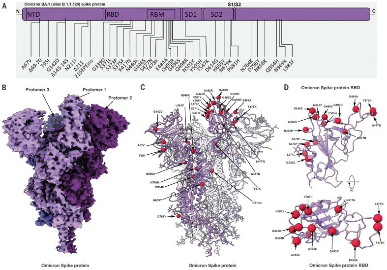
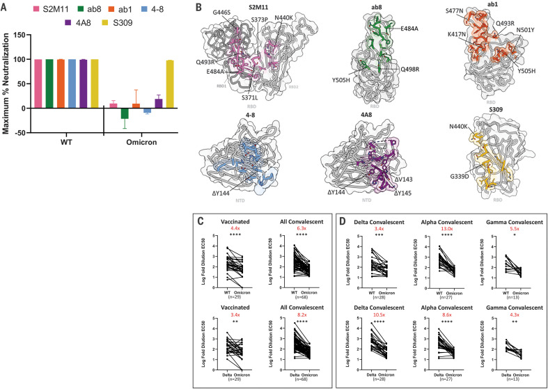
The newly reported Omicron variant is poised to replace Delta as the most prevalent SARS-CoV-2 variant across the world. Cryo-EM structural analysis of the Omicron variant spike protein in complex with human ACE2 reveals new salt bridges and hydrogen bonds formed by mutated residues R493, S496 and R498 in the RBD with ACE2. These interactions appear to compensate for other Omicron mutations such as K417N known to reduce ACE2 binding affinity, resulting in similar biochemical ACE2 binding affinities for Delta and Omicron variants. Neutralization assays show that pseudoviruses displaying the Omicron spike protein exhibit increased antibody evasion. The increase in antibody evasion, together with retention of strong interactions at the ACE2 interface, thus represent important molecular features that likely contribute to the rapid spread of the Omicron variant.


Figures

Fig. 1.
Fig. 1.. Cryo-EM structure of the Omicron spike protein.
(A) A schematic diagram illustrating the domain arrangement of the spike protein. Mutations present in the Omicron variant spike protein are labeled. RBM, receptor binding motif. (B) Cryo-EM map of the Omicron spike protein at 2.79-Å resolution. Protomers are colored in different shades of purple. (C) Cryo-EM structure of Omicron spike protein indicating the locations of modeled mutations on one protomer. (D) The Omicron spike protein RBD shown in two orthogonal orientations with Cα positions of the mutated residues shown as red spheres. Single-letter abbreviations for the amino acid residues are as follows: A, Ala; C, Cys; D, Asp; E, Glu; F, Phe; G, Gly; H, His; I, Ile; K, Lys; L, Leu; M, Met; N, Asn; P, Pro; Q, Gln; R, Arg; S, Ser; T, Thr; V, Val; W, Trp; and Y, Tyr.
Fig. 2.
Fig. 2.. SPR analysis of the wild-type, Delta, and Omicron spike protein affinities for human ACE2.
(A to C) Representative traces of single-cycle kinetic analyses of spike protein–ACE2 binding. The raw data (black) is fit (red) to a model using a 1:1 binding stoichiometry from which apparent dissociation constants were derived. The curves were obtained by injecting 6.25, 31.25, 62.5, 125, and 250 nM of each spike protein in successive cycles. RU, response units; WT, wild type. (D) Quantitation of apparent dissociation constants (KD,app) for the wild-type, Delta, and Omicron spike protein–ACE2 interactions. The standard deviation obtained from at least three technical replicates is shown. Horizontal dotted lines are plotted for mutants carrying only K417N (top) or N501Y and E484K (Glu484→Lys; bottom) mutations to demonstrate the range of this assay (see fig. S2 for binding data). A Tukey’s multiple comparisons test was performed on the wild-type, Delta, and Omicron binding affinities (*P ≤ 0.05; ns, not significant). A table highlighting the fold changes in KD,app for the Delta and Omicron spike protein–ACE2 interactions relative to wild type is shown.
Fig. 3.
Fig. 3.. Cryo-EM structure of the Omicron spike protein–ACE2 complex.
(A) Cryo-EM map of the Omicron spike protein in complex with human ACE2 at 2.45-Å resolution after global refinement. The three protomers are colored in different shades of purple, and the density for bound ACE2 is colored in blue. (B) Cryo-EM map of the Omicron spike protein RBD in complex with ACE2 at 2.66-Å resolution after focused refinement. The boxed area indicates the region highlighted in (C). (C) Cryo-EM density mesh at the Omicron spike protein RBD–ACE2 interface, with fitted atomic model. Yellow and red dashed lines represent new hydrogen bonds and ionic interactions, respectively. (D to F) Comparison of the RBD-ACE2 interface between the Omicron (top) and Delta (bottom) variants. Compared with the Delta variant, new interactions are formed as a result of the mutations Q493R, G496S, and Q498R (D) and local structural changes owing to the N501Y and Y505H (Tyr505→His) mutations (E) present in the Omicron variant. The salt bridge between Delta RBD K417 and ACE2 D30 that is present in the Delta variant spike protein but lost in the Omicron variant is highlighted in (F). Yellow and red dashed lines represent hydrogen bonds and ionic interactions, respectively.
Fig. 4.
Fig. 4.. Monoclonal antibodies and vaccinated and convalescent patient-derived sera exhibit decreased Omicron neutralization potency.
(A) Maximum neutralization achieved by the indicated monoclonal antibodies against wild-type and Omicron pseudoviruses (n = 3 technical replicates). Error bars denote standard deviation of the mean. (B) Antibody binding footprints for the monoclonal antibodies tested in this study. Omicron spike protein mutations that fall within each antibody footprint are labeled. (C) Log-fold 50% effective concentration (EC50) dilutions for vaccinated and convalescent patient sera for either wild-type (WT) versus Omicron variant pseudoviruses (top) or Delta versus Omicron variant pseudoviruses (bottom). (D) As in (C) but with a breakdown of the convalescent patients into previous infection with Delta, Alpha, or Gamma variants of concern. A pairwise statistical significance test was performed using the Wilcoxon matched pairs test (*P ≤ 0.05; **P ≤ 0.01; ***P ≤ 0.001; ****P ≤ 0.0001). The fold change in the geometric mean between the two groups is shown in red at the top of each plot.

Comment in

References

    1. Elbe S., Buckland-Merrett G., Data, disease and diplomacy: GISAID’s innovative contribution to global health. Glob. Chall. 1, 33–46 (2017). 10.1002/gch2.1018 - DOI - PMC - PubMed
    1. Hoffmann M., Kleine-Weber H., Schroeder S., Krüger N., Herrler T., Erichsen S., Schiergens T. S., Herrler G., Wu N.-H., Nitsche A., Müller M. A., Drosten C., Pöhlmann S., SARS-CoV-2 cell entry depends on ACE2 and TMPRSS2 and is blocked by a clinically proven protease inhibitor. Cell 181, 271–280.e8 (2020). 10.1016/j.cell.2020.02.052 - DOI - PMC - PubMed
    1. Piccoli L., Park Y.-J., Tortorici M. A., Czudnochowski N., Walls A. C., Beltramello M., Silacci-Fregni C., Pinto D., Rosen L. E., Bowen J. E., Acton O. J., Jaconi S., Guarino B., Minola A., Zatta F., Sprugasci N., Bassi J., Peter A., De Marco A., Nix J. C., Mele F., Jovic S., Rodriguez B. F., Gupta S. V., Jin F., Piumatti G., Lo Presti G., Pellanda A. F., Biggiogero M., Tarkowski M., Pizzuto M. S., Cameroni E., Havenar-Daughton C., Smithey M., Hong D., Lepori V., Albanese E., Ceschi A., Bernasconi E., Elzi L., Ferrari P., Garzoni C., Riva A., Snell G., Sallusto F., Fink K., Virgin H. W., Lanzavecchia A., Corti D., Veesler D., Mapping neutralizing and immunodominant sites on the SARS-CoV-2 spike receptor-binding domain by structure-guided high-resolution serology. Cell 183, 1024–1042.e21 (2020). 10.1016/j.cell.2020.09.037 - DOI - PMC - PubMed
    1. Wei C., Shan K.-J., Wang W., Zhang S., Huan Q., Qian W., Evidence for a mouse origin of the SARS-CoV-2 Omicron variant. J. Genet. Genomics 10.1016/j.jgg.2021.12.003 (2021). 10.1016/j.jgg.2021.12.003 - DOI - PMC - PubMed
    1. Walls A. C., Park Y.-J., Tortorici M. A., Wall A., McGuire A. T., Veesler D., Structure, function, and antigenicity of the SARS-CoV-2 spike glycoprotein. Cell 181, 281–292.e6 (2020). 10.1016/j.cell.2020.02.058 - DOI - PMC - PubMed

Publication types

MeSH terms

Supplementary concepts