Review
. 2022 Jan 21;130(2):273-287.
doi: 10.1161/CIRCRESAHA.121.319810. Epub 2022 Jan 20.

Detecting Cardiovascular Protein-Protein Interactions by Proximity Proteomics

Affiliations
Review

Detecting Cardiovascular Protein-Protein Interactions by Proximity Proteomics

Jared S Kushner et al. Circ Res. .

Abstract

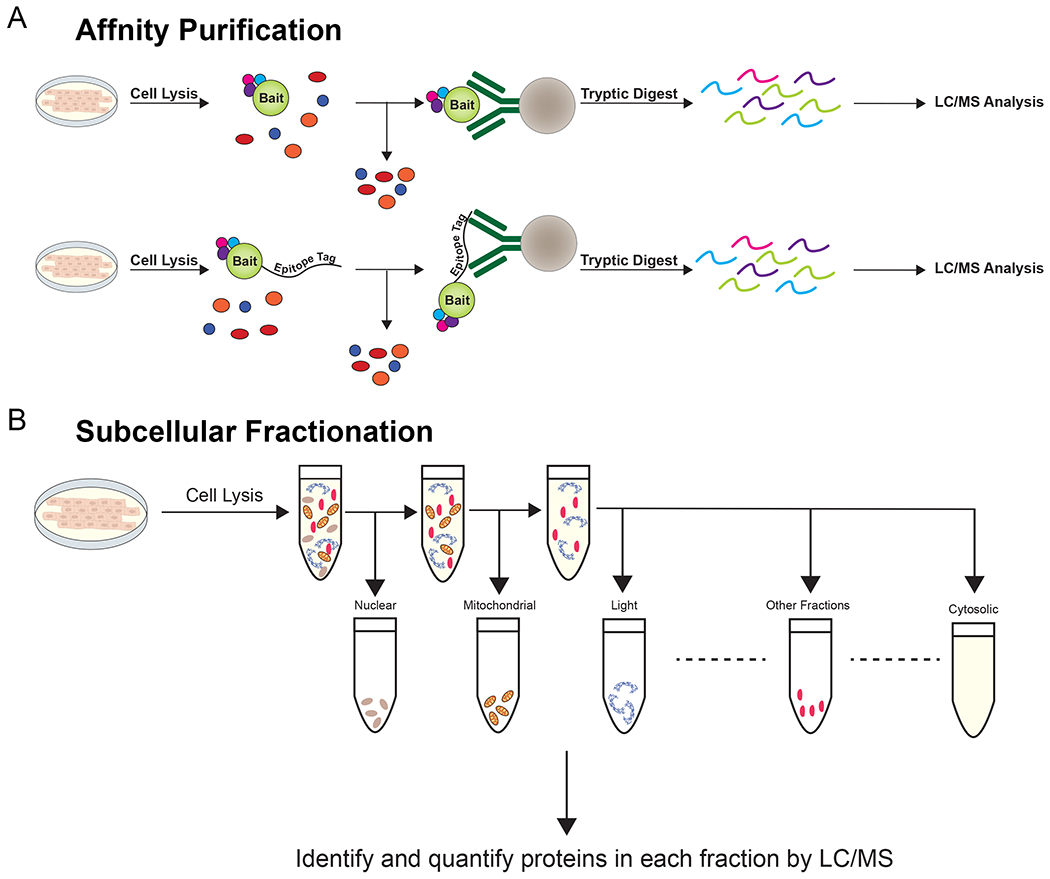
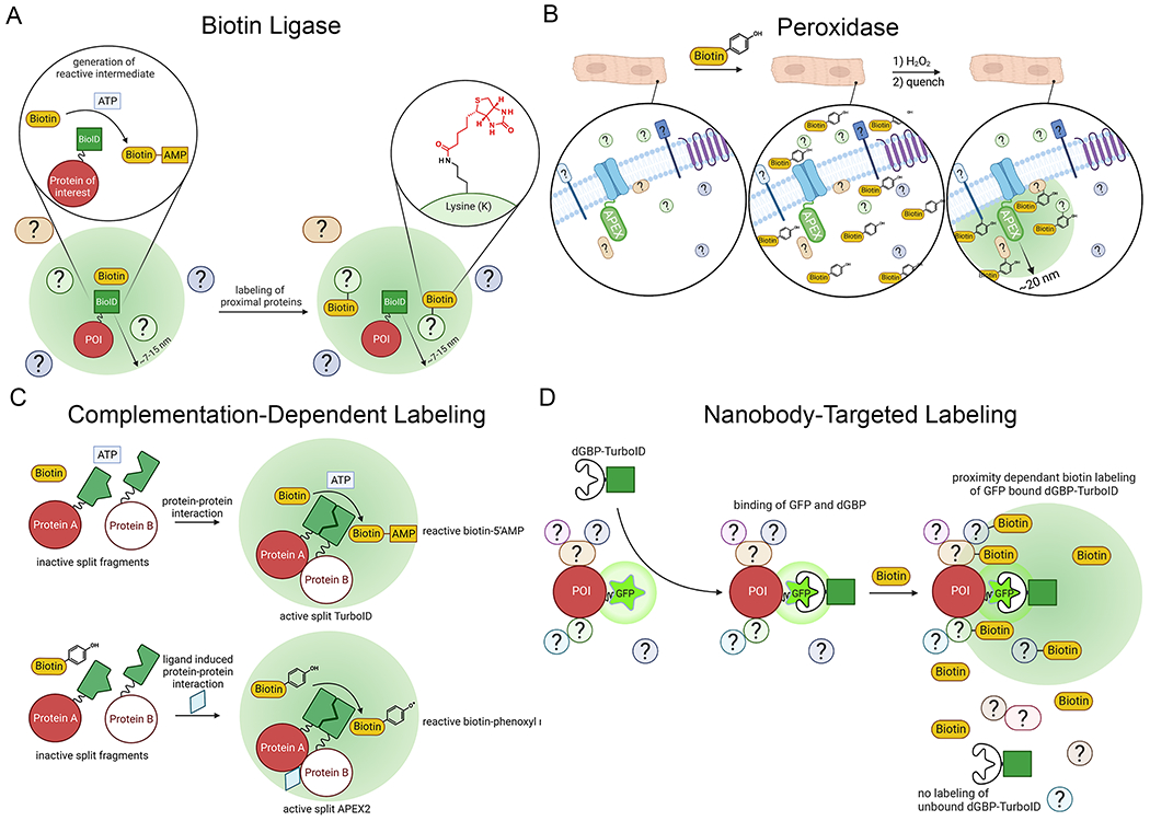
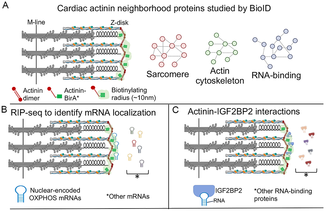
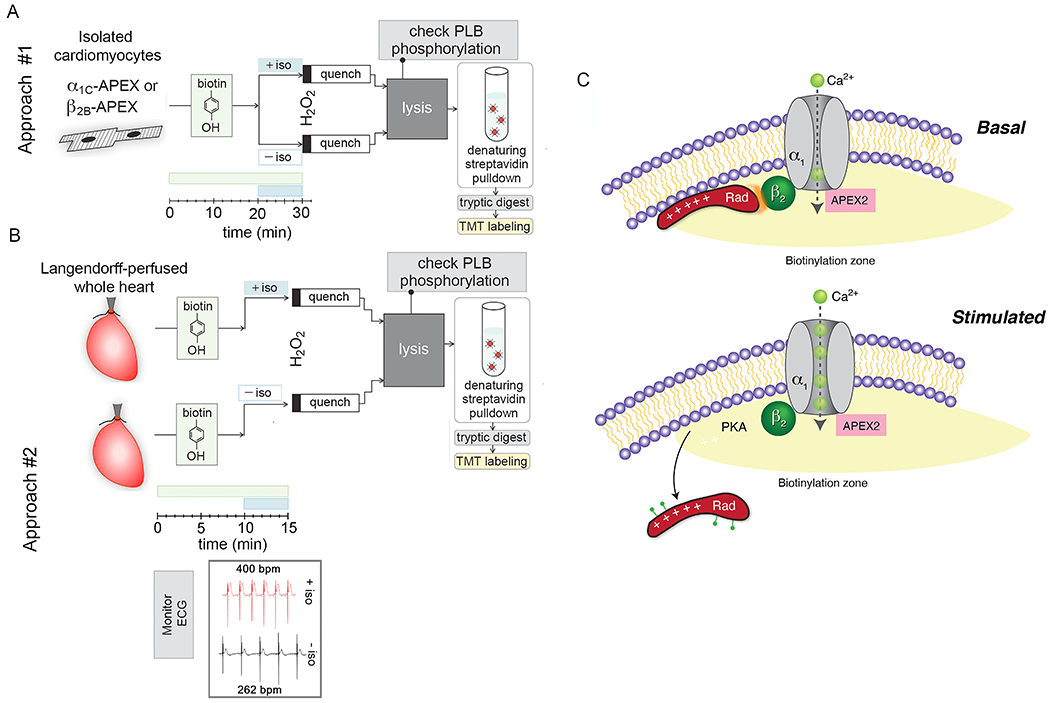
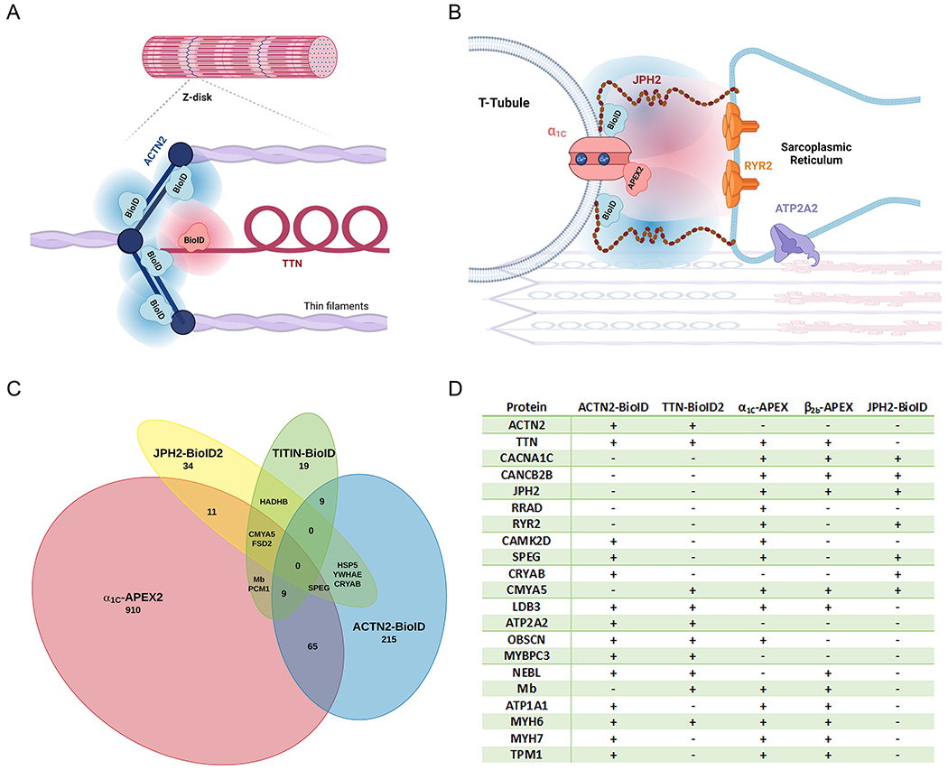
Rapidly changing and transient protein-protein interactions regulate dynamic cellular processes in the cardiovascular system. Traditional methods, including affinity purification and mass spectrometry, have revealed many macromolecular complexes in cardiomyocytes and the vasculature. Yet these methods often fail to identify in vivo or transient protein-protein interactions. To capture these interactions in living cells and animals with subsequent mass spectrometry identification, enzyme-catalyzed proximity labeling techniques have been developed in the past decade. Although the application of this methodology to cardiovascular research is still in its infancy, the field is developing rapidly, and the promise is substantial. In this review, we outline important concepts and discuss how proximity proteomics has been applied to study physiological and pathophysiological processes relevant to the cardiovascular system.

Keywords: animals; mass spectrometry; muscle cells; protein interaction mapping; proteomics.


Conflict of interest statement

Competing Interests

The authors declare no competing interests

Figures

Figure 1.
Figure 1.. Standard methods for identifying protein-protein interactions.
(A) In affinity purification, cell lysates are exposed to antibodies against the bait protein. These antibodies are precipitated and interacting proteins are digested and identified by LC/MS. To study interactors of bait proteins with universal epitope-tag directed antibodies, the fusion of a standard epitope tag can overcome the need for protein-specific antibodies. (B) In lysates subjected to sucrose gradient centrifugation, organelles and subcellular locations of interest are enriched based on their densities.
Figure 2.
Figure 2.. Proteomic mapping by proximity labeling.
(A) In an ATP dependent process, biotin ligases covalently modify basic residues such as lysine with biotin. (B) In cells loaded with biotin-phenol and treated with H2O2, peroxidases generate biotin-phenoxy radicals which attack nearby electron-rich amino acids, preferentially tyrosine. The labeling radius is determined by the longevity of these radicals prior to their quenching. (C) Cleaved or Split-TurboID and Split-APEX2 systems have been developed such that complementation and subsequent biotin-phenol/biotin dependent-proximity labeling occurs when the cleaved enzyme fusions are close together , . This complementation increases labeling specificity for the cellular domain where the fused proteins interact. (D) In this modular labeling system, a nanobody to GFP that is destabilized when not bound to GFP was conjugated to TurboID (dGFP-TurboID), which is co-expressed with a GFP-fused protein of interest (POI) . The destabilization of the nanobody ensures high labeling specificity for the POI. Illustration Credit: Ben Smith.
Figure 3.
Figure 3.. Use of BioID to probe the cardiac actinin neighborhood.
(A) Actinin-BirA* was expressed, via CRISPR knock-in, in human inducible pluripotent stem cell derived cardiomyocytes. Biotinylated proteins were purified and identified by mass spectrometry. Gene ontology analysis of the proteins revealed functions in actin cytoskeleton, sarcomere, and RNA binding. The light-green shading indicates an estimate of the zone of biotinylation. (B) RNA precipitation followed by sequencing (RIP-seq) using streptavidin affinity purification to pull-down RNA-binding proteins that were biotinylated within ~10 nm of actinin-BirA*. Gene ontology analysis indicated enrichment of oxidative phosphorylation (OXPHOS) transcripts. (C) Actinin interacts directly with insulin-like growth factor 2 mRNA binding protein 2 (IGF2BP2), localizing IGF2BP2 to Z-disks. Adapted from .
Figure 4.
Figure 4.. APEX2-based proximity labeling identifies mechanism underlying adrenergic regulation of CaV1.2 in heart.
(A-B) Workflow for processing proximity-labeling in isolated cardiomyocytes and retrograde-perfused hearts. V5 epitope and APEX2 cDNA were conjugated to the N-terminus of dihydropyridine-resistant α1C and wild-type β2B subunits. Transgenic mice with non-targeted insertion of tetracycline-regulated cDNAs were bred with cardiac-specific codon-optimized reverse transcriptional transactivator (rtTA) mice . Transgene expression did not require doxycycline due to low basal binding of rtTA to the Tet operator sequences. In approach #1, ventricular myocytes were isolated by enzymatic digestion and proximity labeling was performed as previously described for isolated cells except for the addition of 1 μM isoproterenol for 10 minutes prior to labeling with H2O2. For each heart, samples were split into 2 groups, one with isoproterenol and one without isoproterenol. After lysis, the effect of PKA on phospholamban (PLB) was determined by western blotting to ensure viability of cardiomyocytes during labeling. In approach #2 (B), samples could not be paired, and hearts were labeled in the absence or presence of isoproterenol. Incubation with biotin-phenol was reduced to 15 minutes and isoproterenol for 5 minutes. Bpm= beats per minutes. (C) Schematic demonstrating zone of biotinylation around CaV1.2 channels. PKA is recruited to and Rad is depleted from the channel neighborhood after isoproterenol. Adapted from .
Figure 5:
Figure 5:. Proximity proteomes of the cardiac Z-disk and cardiac dyad.
(A-B) Schematics illustrating the foci of published proteomic mapping studies located at the Z-disk (A) and the overlying sarcolemma (B). (C) Overlapping proteomic mapping of sarcolemmal and Z-disk protein neighborhoods. Proteins included were described by authors as showing significant enrichment in the neighborhood of their protein of interest. In vivo proximity labeling occurred in knock-in mice with BioID fused to Ttn, BioID2 fused to Jph2, human iPSC-derived cardiac myocytes with knock-in of BioID fused to ACTN2. Live in-situ ex-vivo proximity labeling was examined in mice with inducible transgenic overexpression of the CaV1.2 α1C subunit fused to APEX2. (D) Enrichment of proteins critical for cell structure, Ca2+ homeostasis, contraction and signaling were noted across multiple proximity proteomes, including those from isolated myocytes from mice overexpressing CaV1.2 β2B-APEX2. Created in part with LucidChart and Biorender.com , , , .

References

    1. Agou F, Veron M. In vivo protein cross-linking. Methods Mol Biol. 2015;1278:391–405 - PubMed
    1. Christopher JA, Stadler C, Martin CE, Morgenstern M, Pan Y, Betsinger CN, Rattray DG, Mahdessian D, Gingras AC, Warscheid B, et al. Subcellular proteomics. Nat Rev Methods Primers. 2021;1 - PMC - PubMed
    1. Mellacheruvu D, Wright Z, Couzens AL, Lambert JP, St-Denis NA, Li T, Miteva YV, Hauri S, Sardiu ME, Low TY, et al. The crapome: A contaminant repository for affinity purification-mass spectrometry data. Nat Methods. 2013;10:730–736 - PMC - PubMed
    1. Choi H, Liu G, Mellacheruvu D, Tyers M, Gingras AC, Nesvizhskii AI. Analyzing protein-protein interactions from affinity purification-mass spectrometry data with saint. Curr Protoc Bioinformatics. 2012;Chapter 8:Unit8 15. - PMC - PubMed
    1. Roux KJ, Kim DI, Raida M, Burke B. A promiscuous biotin ligase fusion protein identifies proximal and interacting proteins in mammalian cells. J Cell Biol. 2012;196:801–810 - PMC - PubMed

Publication types