. 2021 Dec 23;14(1):14.
doi: 10.3390/toxins14010014.

Structural Analysis of Botulinum Neurotoxins Type B and E by Cryo-EM

Affiliations

Structural Analysis of Botulinum Neurotoxins Type B and E by Cryo-EM

Sara Košenina et al. Toxins (Basel). .

Abstract

Botulinum neurotoxins (BoNTs) are the causative agents of a potentially lethal paralytic disease targeting cholinergic nerve terminals. Multiple BoNT serotypes exist, with types A, B and E being the main cause of human botulism. Their extreme toxicity has been exploited for cosmetic and therapeutic uses to treat a wide range of neuromuscular disorders. Although naturally occurring BoNT types share a common end effect, their activity varies significantly based on the neuronal cell-surface receptors and intracellular SNARE substrates they target. These properties are the result of structural variations that have traditionally been studied using biophysical methods such as X-ray crystallography. Here, we determined the first structures of botulinum neurotoxins using single-particle cryogenic electron microscopy. The maps obtained at 3.6 and 3.7 Å for BoNT/B and /E, respectively, highlight the subtle structural dynamism between domains, and of the binding domain in particular. This study demonstrates how the recent advances made in the field of single-particle electron microscopy can be applied to bacterial toxins of clinical relevance and the botulinum neurotoxin family in particular.

Keywords: BoNT/B; BoNT/E; Clostridium botulinum; botulinum neurotoxin; botulism; cryo-EM.


Conflict of interest statement

The authors declare no conflict of interest. The funders had no role in the design of the study; in the collection, analyses, nor interpretation of data; in the writing of the manuscript; nor in the decision to publish the results.

Figures

Figure 1
Figure 1
(a) Phylogenetic tree of clostridial neurotoxins prepared with MEGAX [33] from protein sequences aligned with CLUSTALO [34]. (b) Schematic representation of the botulinum neurotoxin domain structure. The toxins are expressed as single-chain proteins but are later proteolytically converted to their active di-chain form, where LC is linked by a single disulphide bridge to HC. The LHN fragment has been defined previously as the combined LC + HN domains [35].
Figure 2
Figure 2
(a,b) Cryo-EM maps of BoNT/B and BoNT/E at an estimated resolution of 3.6 and 3.7 Å, respectively. The maps, as well as comparative 2D class averages of the toxins in (c,d), reveal clear secondary elements and domain arrangements illustrated by a schematic diagram (LC in green, HN in blue, HC in grey).
Figure 3
Figure 3
(a,b) 3D reconstruction of BoNT/B (teal) and BoNT/E (red), respectively. (c,d) Close-up map of secondary features from each domain, including the mutated catalytic site (mutations marked by *, E231Q/H234Y and E213Q/H216Y for BoNT/B and BoNT/E, respectively); α-helix of HN, β-sheet of HCN, and the ganglioside-binding site of HCC.
Figure 4
Figure 4
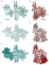
(a) Superposition of the cryo-EM (teal) and crystal (PDB 1EPW, light grey) coordinates of BoNT/B. (b,c) B factor-scaled ribbon representations of the crystal and cryo-EM structures of BoNT/B, respectively. (d) Superposition of the cryo-EM (red) and crystal (PDB 3FFZ, light pink) coordinates of BoNT/E. (e,f) B factor-scaled ribbon representations of the crystal and cryo-EM structures of BoNT/E, respectively.

References

    1. Williamson C.H., Sahl J.W., Smith T.J., Xie G., Foley B.T., Smith L.A., Fernández R.A., Lindström M., Korkeala H., Keim P., et al. Comparative genomic analyses reveal broad diversity in botulinum-toxin-producing Clostridia. BMC Genom. 2016;17:180. doi: 10.1186/s12864-016-2502-z. - DOI - PMC - PubMed
    1. Zhang S., Masuyer G., Zhang J., Shen Y., Lundin D., Henriksson L., Miyashita S.I., Martinez-Carranza M., Dong M., Stenmark P. Identification and characterization of a novel botulinum neurotoxin. Nat. Commun. 2017;8:14130. doi: 10.1038/ncomms14130. - DOI - PMC - PubMed
    1. Dong M., Masuyer G., Stenmark P. Botulinum and tetanus neurotoxins. Annu. Rev. Biochem. 2019;88:811–837. doi: 10.1146/annurev-biochem-013118-111654. - DOI - PMC - PubMed
    1. Rossetto O., Montecucco C. Tables of toxicity of botulinum and tetanus neurotoxins. Toxins. 2019;11:686. doi: 10.3390/toxins11120686. - DOI - PMC - PubMed
    1. Rasetti-Escargueil C., Popoff M.R. Antibodies and vaccines against botulinum toxins: Available measures and novel approaches. Toxins. 2019;11:528. doi: 10.3390/toxins11090528. - DOI - PMC - PubMed

Publication types

MeSH terms