. 2022 Jan 11;9(1):26.
doi: 10.3390/vetsci9010026.

Feline and Canine Cutaneous Lymphocytosis: Reactive Process or Indolent Neoplastic Disease?

Affiliations

Feline and Canine Cutaneous Lymphocytosis: Reactive Process or Indolent Neoplastic Disease?

Francesco Albanese et al. Vet Sci. .

Abstract

Cutaneous lymphocytosis (CL) is an uncommon and controversial lymphoproliferative disorder described in dogs and cats. CL is generally characterized by a heterogeneous clinical presentation and histological features that may overlap with epitheliotropic lymphoma. Therefore, its neoplastic or reactive nature is still debated. Here, we describe clinicopathological, immunohistochemical, and clonality features of a retrospective case series of 19 cats and 10 dogs with lesions histologically compatible with CL. In both species, alopecia, erythema, and scales were the most frequent clinical signs. Histologically, a dermal infiltrate of small to medium-sized lymphocytes, occasionally extending to the subcutis, was always identified. Conversely, when present, epitheliotropism was generally mild. In cats, the infiltrate was consistently CD3+; in dogs, a mixture of CD3+ and CD20+ lymphocytes was observed only in 4 cases. The infiltrate was polyclonal in all cats, while BCR and TCR clonal rearrangements were identified in dogs. Overall, cats had a long-term survival (median overall survival = 1080 days) regardless of the treatment received, while dogs showed a shorter and variable clinical course, with no evident associations with clinicopathological features. In conclusion, our results support a reactive nature of the disease in cats, associated with prolonged survival; despite a similar histological picture, canine CL is associated with a more heterogeneous lymphocytic infiltrate, clonality results, and response to treatment, implying a more challenging discrimination between CL and CEL in this species. A complete diagnostic workup and detailed follow-up information on a higher number of cases is warrant for dogs.

Keywords: PARR; cat; cutaneous lymphocytosis; dog; immunohistochemistry; lymphoma; skin.


Conflict of interest statement

The authors declare no conflict of interest. The funders had no role in the design of the study; in the collection, analyses, or interpretation of data; in the writing of the manuscript; or in the decision to publish the results.

Figures

Figure 1
Figure 1
Clinical signs in cats. (a) Case no. 3: generalized alopecia and scales (courtesy Dr. Fabrizio Fabbrini). (b) Case no. 4: focal alopecia with multifocal ulcers on the abdomen (courtesy Dr. Alessandro Corona). (c) Case no. 3: erythema, alopecia, and scales on both right eyelids (courtesy Dr. Sivia Colombo). (d) Case no. 14: alopecia, erythema, and swelling of the nose and eyelids (courtesy of Dr. Ilaria Mannara).
Figure 2
Figure 2
Clinical signs in dogs. (a) Case no. 4: severe generalized alopecia with erythematous and exfoliative dermatitis in a Poodle (courtesy Dr. Roberta Gamba). (b) Case no. 2: erythematous plaques on the inner surface of the pinna (courtesy Dr. Claudia d’Angeli). (c) Case no. 1: multifocal erythematous irregular plaques on the abdomen (courtesy Dr. Francesco Carrani). (d) Case no. 6: alopecia, erythema, small yellowish scales on the neck (courtesy Dr. Francesca Carraro).
Figure 3
Figure 3
Histological findings in cases of feline and canine cutaneous lymphocytosis (H&E stain). (a) Cat no. 3: diffuse lymphocytic infiltrates in the superficial dermis and panniculus, note lymphocyte aggregates in the panniculus (20×). (b) Dog no. 6: diffuse lymphocytic infiltrate in the superficial dermis (20×). (c) Cat no. 8: mild epitheliotropism by small lymphocyte aggregates (asterisks) (400×). (d) Dog no. 4: mild epitheliotropism by single lymphocytes (asterisks) (400×). (e) Cat no. 3: diffuse infiltrate of small lymphocytes with few infiltrating eosinophils (black arrows in the figure and in the inset) (400×). (f) Cat no. 7: diffuse infiltrate of small lymphocytes with few infiltrating mast cells (white arrow in the figure and in the inset) and eosinophils (black arrows in the figure and in the inset) (400×).
Figure 4
Figure 4
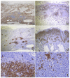
Immunohistochemical findings of feline and canine lymphocytosis (peroxidase method). (a) Cat no. 3: infiltrating cells are diffusely positive with the anti-CD3 antibody (40×). (b) Dog no. 8: infiltrating cells are diffusely positive with the anti-CD3 antibody (40×). (c) Cat no. 3: deep nodular aggregates of lymphocytes (asterisks) are positive for the anti-CD20 antibody (B lymphocytes) (40×). (d) Dog no. 6: infiltrating cells are diffusely positive with the anti-CD20 antibody for B lymphocytes (40×). (e) Cat no. 6: lymphocytes are diffusely positive for anti-CD3 antibody, note the scant epitheliotropism (black arrows, 400×). (f) Dog no. 3: CD3+ T lymphocytes are present, single or in small aggregates (400×).
Figure 5
Figure 5
Clinical follow up of dog no. 9. (a) The dog at presentation: severe generalized erythematous and exfoliative dermatitis. (b) The dog at 11-month follow-up after removal of all drugs: relevant improvement of clinical signs with almost complete hair regrowth (courtesy Dr. Roberto Damiani).
Figure 6
Figure 6
Suggested clinical and diagnostic algorithm in suspected cases of cutaneous lymphocytosis.

Similar articles

Cited by

References

    1. Ploysangam T., Breneman D.L., Mutasim D.F. Cutaneous pseudolymphomas. J. Am. Acad. Dermatol. 1998;38:877–898. doi: 10.1016/S0190-9622(98)70154-9. - DOI - PubMed
    1. Bergman R. Pseudolymphoma and cutaneous lymphoma: Facts and controversies. Clin. Dermatol. 2010;28:568–574. doi: 10.1016/j.clindermatol.2010.04.005. - DOI - PubMed
    1. Gilliam A.C., Wood G.S. Cutaneous lymphoid hyperplasias. Semin. Cutan. Med. Surg. 2000;19:133–141. doi: 10.1053/sder.2000.8329. - DOI - PubMed
    1. Gilbert S., Affolter V.K., Gross T.L., Moore P.F., Ihrke P.J. Clinical, morphological and immunohistochemical characterization of cutaneous lymphocytosis in 23 cats. Vet. Dermatol. 2004;15:3–12. doi: 10.1111/j.1365-3164.2004.00352.x. - DOI - PubMed
    1. Affolter V.K., Gross T.L., Moore P.F. Indolent cutaneous T-cell lymphoma presenting as cutaneous lymphocytosis in dogs. Vet. Dermatol. 2009;20:577–585. doi: 10.1111/j.1365-3164.2009.00833.x. - DOI - PubMed

Grants and funding

LinkOut - more resources