

Overexpression of PD-1 and CD39 in tumor-infiltrating lymphocytes compared with peripheral blood lymphocytes in triple-negative breast cancer
- PMID: 35051220
- PMCID: PMC8775239
- DOI: 10.1371/journal.pone.0262650
Overexpression of PD-1 and CD39 in tumor-infiltrating lymphocytes compared with peripheral blood lymphocytes in triple-negative breast cancer
Abstract
Background and aim: Growing evidence highlighted the primary role of the immune system in the disease course of triple-negative breast cancer (TNBC). The study aim was to investigate the expression of PD-1 and CD39 on CD4+ and CD8+ cells infiltrating tumor tissue compared to their counterparts in peripheral blood and explore its association with tumor characteristics, disease progression, and prognosis in females with TNBC.
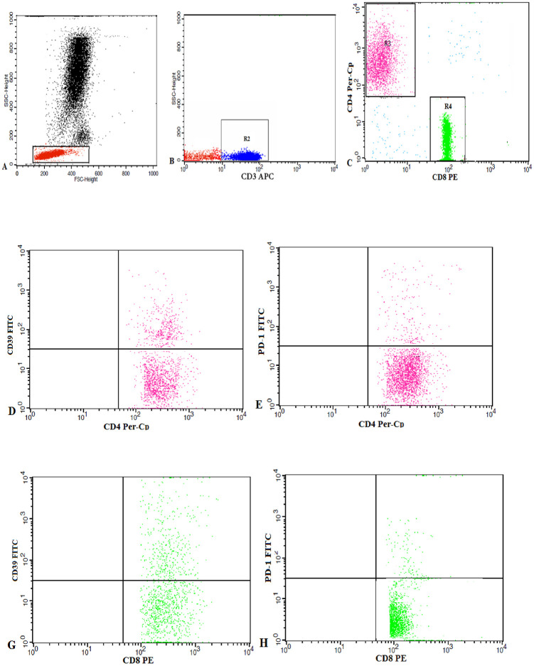
Patients and methods: The study included 30 TNBC patients and 20 healthy controls. Cancer and normal breast tissue and peripheral blood samples were collected for evaluation of the expression of PD-1 and CD39 on CD4+ and CD8+T cells by flow cytometry.
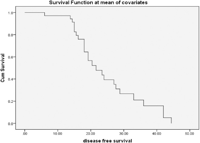
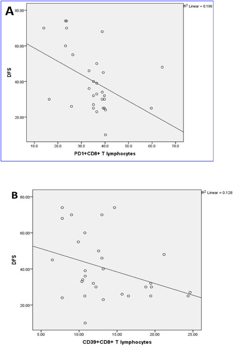
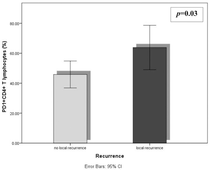
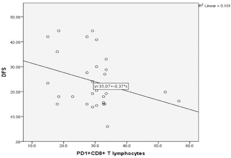
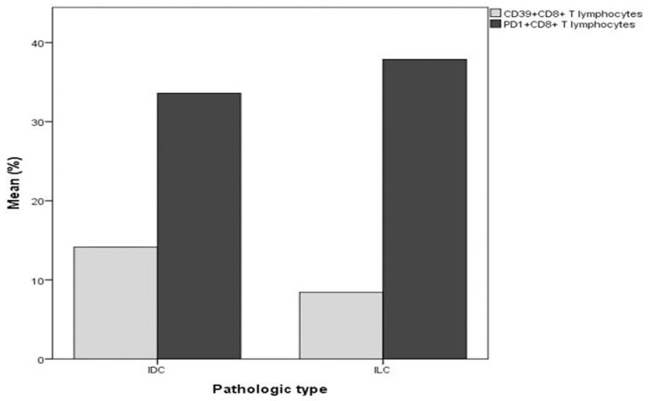
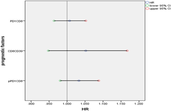
Results: A marked reduction in the percentage of CD8+ T lymphocytes and a significant increase in the frequencies of CD4+ T lymphocytes and CD4+ and CD8+ T lymphocytes expressing PD1 and CD39 were evident in tumor tissue in comparison with the normal breast tissue. The DFS was inversely related to the cancer tissue PD1+CD8+ and CD39+CD8+ T lymphocytes. Almost all studied cells were significantly increased in the tumor tissue than in peripheral blood. Positive correlations were detected among peripheral PD1+CD4+T lymphocytes and each of cancer tissue PD1+CD4+, PD1+CD8+and CD39+CD8+T cells, and among peripheral and cancer tissue CD39+CD4+and CD39+CD8+ T cells.
Conclusions: The CD39 and PD1 inhibitory pathways are synergistically utilized by TNBC cells to evade host immune response causing poor survival. Hence, combinational immunotherapy blocking these pathways might be a promising treatment strategy in this type of cancer.
Conflict of interest statement
the authors have declared that no competing interests exist
Figures
References
-
- Bray F, Ferlay J, Soerjomataram I, Siegel RL, Torre LA, Jemal A. Global cancer statistics 2018: GLOBOCAN estimates of incidence and mortality worldwide for 36 cancers in 185 countries. CA: a cancer journal for clinicians. 2018;68(6):394–424. - PubMed
-
- Miyashita M, Sasano H, Tamaki K, Hirakawa H, Takahashi Y, Nakagawa S, et al.. Prognostic significance of tumor-infiltrating CD8+ and FOXP3+ lymphocytes in residual tumors and alterations in these parameters after neoadjuvant chemotherapy in triple-negative breast cancer: a retrospective multicenter study. Breast Cancer Res. 2015;17(1):124. doi: 10.1186/s13058-015-0632-x - DOI - PMC - PubMed
MeSH terms
Substances
LinkOut - more resources
Full Text Sources
Research Materials
