. 2022 Apr 7;139(14):2227-2239.
doi: 10.1182/blood.2021013113.

CRLF3 plays a key role in the final stage of platelet genesis and is a potential therapeutic target for thrombocythemia

Affiliations

CRLF3 plays a key role in the final stage of platelet genesis and is a potential therapeutic target for thrombocythemia

Cavan Bennett et al. Blood. .

Abstract

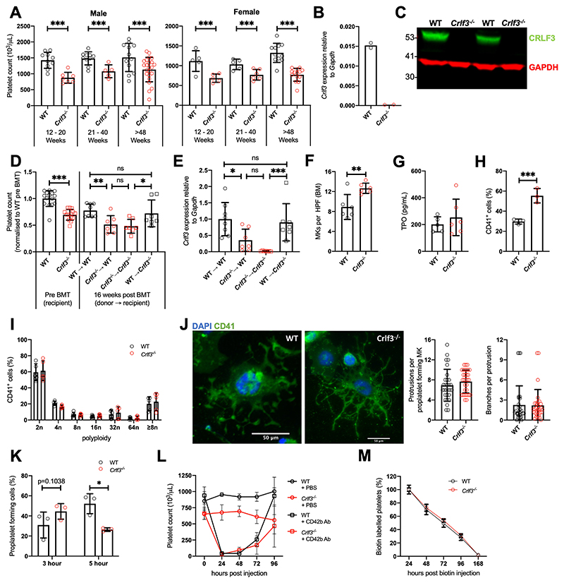
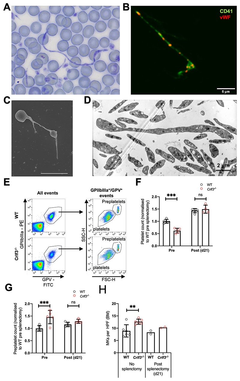
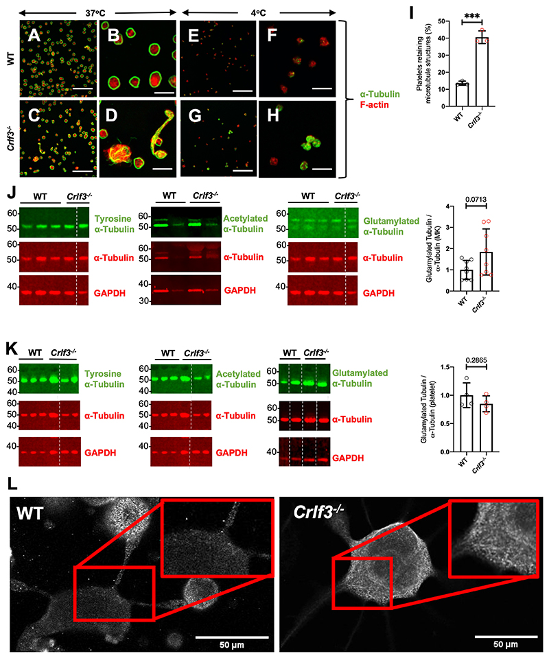
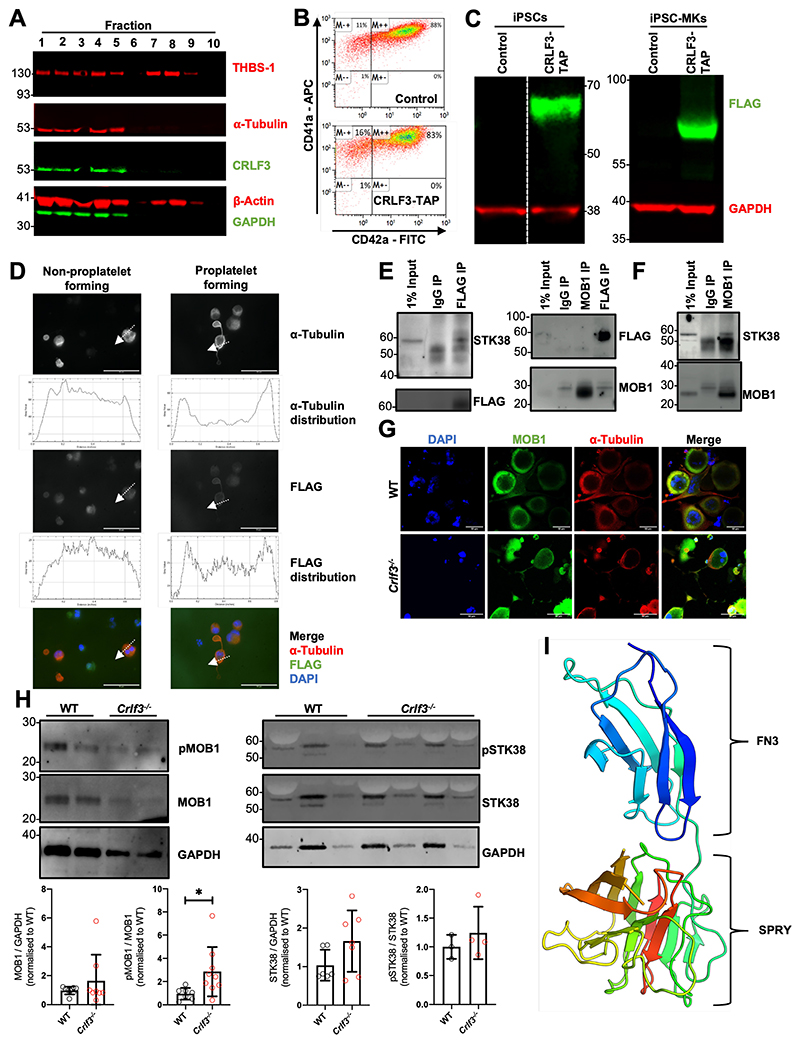
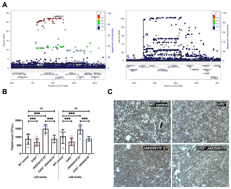
The process of platelet production has so far been understood to be a 2-stage process: megakaryocyte maturation from hematopoietic stem cells followed by proplatelet formation, with each phase regulating the peripheral blood platelet count. Proplatelet formation releases into the bloodstream beads-on-a-string preplatelets, which undergo fission into mature platelets. For the first time, we show that preplatelet maturation is a third, tightly regulated, critical process akin to cytokinesis that regulates platelet count. We show that deficiency in cytokine receptor-like factor 3 (CRLF3) in mice leads to an isolated and sustained 25% to 48% reduction in the platelet count without any effect on other blood cell lineages. We show that Crlf3-/- preplatelets have increased microtubule stability, possibly because of increased microtubule glutamylation via the interaction of CRLF3 with key members of the Hippo pathway. Using a mouse model of JAK2 V617F essential thrombocythemia, we show that a lack of CRLF3 leads to long-term lineage-specific normalization of the platelet count. We thereby postulate that targeting CRLF3 has therapeutic potential for treatment of thrombocythemia.


Conflict of interest statement

Conflict of Interest Disclosure

Parsa Akbari is a full-time employee of Regeneron pharmaceuticals and currently holds options and stock of the company.

Figures

Figure 1
Figure 1. CRLF3 deficiency causes a sustained and isolated reduction in platelet count.
(A) Platelet counts of male (n=5-23) and female (n=5-14) young (12-20 weeks), middle aged (21-40 weeks) and old (>48 weeks) control (WT, black) and Crlf3-/- (red) mice. (B) Expression of Crlf3 relative to Gapdh mRNA determined by qRT-PCR of WT (black) and Crlf3-/- (red) isolated from in vitro cultured MKs (n=2). (C) Western blot of platelet lysates against CRLF3 (green) and GAPDH (red) (n=2). (D) Platelet counts pre- (on the left; n=15 WT/14 Crlf3-/-) and 16 weeks post-bone marrow transplantation (BMT; on the right) of control (WT, circles) and Crlf3-/- (squares) recipient mice that received either WT (black) or Crlf3-/- (red) donor cells (n=8 WT->WT/7 all other groups). (E) Chimaerism was estimated by expression of Crlf3 relative to Gapdh mRNA isolated from in vitro cultured MKs by qRT-PCR (n=8 WT->WT/7 all other groups). (F) Quantification of MKs in H&E stained sections of control (WT, black) and Crlf3-/- (red) tibia (n=6). (G) Thrombopoietin (TPO) concentration determined by ELISA in control (WT, black) and Crlf3-/- (red) plasma (n=5 WT/6 Crlf3-/-). (H) Percentage of CD41+ cells from control (WT, black) and Crlf3-/- (red) in vitro MK cultures (n= 3). (I) Polyploidy of in vitro cultured control (WT, black) and Crlf3-/- (red) MKs analysed by flow cytometry (n=5). (J) Mature in vitro cultured MKs were purified by BSA-gradient, seeded onto fibrinogen coated coverslips and incubated at 37°C for 5 hours to induce proplatelet formation. Fixed samples were stained with CD41 (green) and DAPI (blue), and imaged by fluorescence microscopy. Images are representative for Crlf3-/- and control (WT) proplatelet forming MKs. Scale bars are 50μm. Proplatelet morphology of control (WT, black) and Crlf3-/- (grey) MKs was assessed by blindly quantifying the number of protrusions per proplatelet forming MK and number of branches per protrusion (n=29 WT/31 Crlf3-/-). (K) In vitro cultured MKs were seeded onto fibrinogen coated coverslips and incubated at 37°C for 3 or 5 hours to induce proplatelet formation. After confocal microscopy, percentage of proplatelet forming MKs was determined for control (WT, black) and Crlf3-/- (red) (n=3). At least 460 MKs were counted in each condition. (L) Control (WT, black) and Crlf3-/- (red) animals were injected with PBS (circles) or anti-CD42b (0.6μg/g body weight, squares) and platelet counts determined by automated haemocytometer 0, 24, 48, 72 and 96 hours post injection (n=4 Crlf3-/- + CD42b Ab/3 all other groups). (M) Control (WT, black) and Crlf3-/- (red) mice injected with 1mg NHS-biotin and percentage of CD41+/Ter119-/streptavidin+ platelets was determined by flow cytometry at 24, 48, 72, 96 and 168 hours post injection. Percentage of streptavidin positive platelets at 24 hours represents 100% biotin bound platelets (n=5). Data represents mean ± SD. Unpaired 2-tailed Student’s t test (F, G, H, J) with correction for multiple comparisons using the Holm-Sidak method (A), One-way ANOVA (D, E) or Two-way ANOVA (I, K, L, M) with correction for multiple comparisons using the Holm-Sidak method. *, **, *** and ns denote p<0.05, p<0.01, p<0.005 and non-significant, respectively.
Figure 2
Figure 2. CRLF3 deficiency causes ineffective thrombopoiesis
(A) Romanovsky-stained blood smear from Crlf3-/- mouse whole blood taken at 100x magnification under light microscopy. (B) Crlf3-/- blood smear stained with CD41 (green) and vWF (red) and imaged by confocal microscopy. (C) Washed Crlf3-/- platelets fixed and prepared for scanning or (D) transmission electron microscopy. Scale bars are 5μm (B and C) and 2μm (D). (E) Example flow cytometry plots to determine (F) platelet (GPV+/GPIIbIIIa+ events) and (G) preplatelet (GPV+/GPIIbIIIa+ events with larger forward scatter/side scatter than mature platelets ) counts from control (WT, black) and Crlf3-/- (red) splenectomised mice (n=4 Crlf3-/- post-splenectomy/5 all other groups). (H) Quantification of MKs in H&E stained sections of control (WT, black) and Crlf3-/- (red) tibia of non-splenectomised animals (left hand side) or 21 post splenectomy (right hand side) (n=6 non-splenectomised/2 splenectomised). Data represents mean ± SD (except splenectomised mice in H, where data represents mean). Two-way ANOVA with correction for multiple comparisons using the Holm-Sidak method (F and G), Unpaired 2-tailed Student’s t test (H). **, *** and ns denote p<0.01, p<0.005 and non-significant, respectively.
Figure 3
Figure 3. CRLF3 deficiency causes microtubule hyper-stability
Washed platelets maintained at 37°C (control [WT] - A and B and Crlf3-/- - C and D) or stored at 4°C for 3 hours (control [WT] - E and F and Crlf3-/- - G and H) adhered to poly-L-lysine coated coverslips and stained for α-tubulin (green) and F-actin (red). Scale bars are 20μm (A, C, E, G) and 5 μm (B, D, F, H). (I) Platelets retaining microtubule structures after incubation at 4°C were determined by manual counting of images for control (WT, black) and Crlf3-/- (red) mice (n=3). (J) Representative western blots of in vitro cultured MK or (K) platelet lysates against tyrosine α-tubulin, α-tubulin and GAPDH left panel; acetylated α-tubulin, α-tubulin and GAPDH middle panel; or glutamylated α-tubulin (AG-20B-0020_upper band), α-tubulin and GAPDH right panel for control (WT, black) and Crlf3-/- (red) samples. The quantification of glutamylated α-tubulin/total tubulin (bar graphs on the right) was carried out on 8 control and 8 Crlf3-/- samples with two technical replicates. Where α-tubulin and GAPDH panels are the same for multiple tubulin modifications, membranes were stripped and re-probed between antibodies against specific tubulin modifications before finally being stripped and re-probed for α-tubulin and GAPDH. (L) In vitro cultured MKs were seeded onto fibrinogen coated coverslips and incubated at 37°C for 5 hours to induce proplatelet formation. Samples were fixed, stained for polyglutamylated α-Tubulin (AG-20B-0020) and imaged by fluorescence microscopy. Images are representative for Crlf3-/- and control (WT) proplatelet forming MKs. Scale bars are 50μm. Data represents mean ± SD. Unpaired 2-tailed Student’s (I and K) or Welch’s (J) t test. * and *** denote p<0.05 and p<0.005, respectively.
Figure 4
Figure 4. CRLF3 interacts with the Hippo pathway
(A) Western blot of sucrose gradient centrifugation fractionated human platelets probed with antibodies against α-tubulin, β-actin, thrombospondin (THBS-1), GAPDH and CRLF3. Fractions 1-5 represent cytoskeletal proteins (enriched for α-tubulin and β-actin), whereas fractions 7 and 8 represent granular proteins (enriched for the THBS-1). (B) Representative flow cytometry plots of CRLF3-TAP tagged and control forward programmed iPSC-MKs stained with CD41a and CD42a. (C) Western blot of CRLF3-TAP tagged and control iPSCs and iPSC-MKs probed with antibodies against FLAG (green) and GAPDH (red). (D) CRLF3-TAG tagged iPSC-MKs were seeded onto fibrinogen coated coverslips and incubated at 37°C for 24 hours to induce proplatelet formation. Samples were fixed, stained with α-tubulin (red), FLAG (green) and DAPI (blue), and imaged by fluorescence microscopy. Sub-cellular distribution of α-tubulin and FLAG staining in round and proplatelet forming CRLF3-TAP iPSC-MKs was determined across a section of the MKs along the indicated arrow using ImageJ. Scale bars are 10μm. (E) CRLF3-TAP and (F) control iPSC-MKs were lysed and immunoprecipitated with antibodies against FLAG, MOB1 and IgG. Precipitated lysates were then probed for STK38, MOB1 and FLAG by western blot. (G) In vitro cultured MKs were seeded onto fibrinogen coated coverslips and incubated at 37°C for 5 hours to induce proplatelet formation. Samples were fixed, stained for MOB1, α-Tubulin and DAPI and imaged by fluorescence microscopy. Images are representative for Crlf3-/- and control (WT) proplatelet forming MKs. (H) Western blot of in vitro cultured MKs probed with antibodies against pMOB1, MOB1 and GAPDH (left panel; n=8 MOB1/GAPGH and 4 pMOB1/GAPDH) and pSTK38, STK38 and GAPDH (right panel; n=3 STK38/GAPDH, 3 Crlf3-/- and 4 WT pSTK38/GAPDH). (I) 3D structure of CRLF3 construct 3 (residue 174 to end) solved by experimental phasing. Domains are labelled. Molecular graphics prepared using PyMOL. FN3 = fibronectin type 3. Data represents mean ± SD. Unpaired 2-tailed Student’s t test. * denotes p<0.05.
Figure 5
Figure 5. CRLF3 regulates platelet traits in humans and is a therapeutic target for Essential Thrombocythaemia
(A) Locuszoom of CRLF3 (left) and STK38 (right) showing variants associated with Platelet Distribution Width (PDW) and Mean Platelet Volume (MPV), respectively. The conditionally independent variant is indicated by a purple diamond, LD values (r2) with this variant are indicated by dot colours according to the legend above. The CRLF3 locuszoom plot shows the conditionally independent variant (rs6505211, -log10P: 27.1, MAF: 17.6%) is in high LD with a number of variants which are significantly associated with PDW. In the case of STK38 the locuszoom plot indicates that the conditionally independent variant (rs141301223 -log10P: 10.4, MAF: 0.041%) is not in high LD with nearby variants (common for rare variant associations). (B) Platelet counts from young (≤20-week-old) and old (≥48-week-old) female WT control (black; n=6 young/5 old), Crlf3-/- (red; n=7 young/7 old), JAK2V617F ET (purple; n=7 young/9 old) and Crlf3-/- JAK2V617F (blue; n=6 young/6 old) mice. (C) Fixed tibia sections stained with Gömöri’s reticulin silver stain and imaged by lightX microscopy at 20x magnification. Images are representative of 3 mice per genotype. Data represents mean ± SD. Two-way ANOVA with correction for multiple comparisons using the Holm-Sidak method. *** and ns denote p<0.005 and not significant, respectively.

Comment in

  • The triple crown of platelet generation.
    Becker IC, Italiano JE. Becker IC, et al. Blood. 2022 Apr 7;139(14):2100-2101. doi: 10.1182/blood.2021015018. Blood. 2022. PMID: 35389441 Free PMC article. No abstract available.

References

    1. Zhang L, Orban M, Lorenz M, et al. A novel role of sphingosine 1-phosphate receptor S1pr1 in mouse thrombopoiesis. J Exp Med. 2012;209(12):2165–2181. - PMC - PubMed
    1. Junt T, Schulze H, Chen Z, et al. Dynamic visualization of thrombopoiesis within bone marrow. Science. 2007;317(5845):1767–1770. - PubMed
    1. Thon JN, Montalvo A, Patel-Hett S, et al. Cytoskeletal mechanics of proplatelet maturation and platelet release. J Cell Biol. 2010;191(4):861–874. - PMC - PubMed
    1. Kunishima S, Nishimura S, Suzuki H, Imaizumi M, Saito H. TUBB1 mutation disrupting microtubule assembly impairs proplatelet formation and results in congenital macrothrombocytopenia. Eur J Haematol. 2014;92(4):276–282. - PubMed
    1. Pecci A, Klersy C, Gresele P, et al. MYH9-related disease: a novel prognostic model to predict the clinical evolution of the disease based on genotype-phenotype correlations. Hum Mutat. 2014;35(2):236–247. - PMC - PubMed

Publication types