Review
. 2022 Jan 13;13(1):138.
doi: 10.3390/genes13010138.

Wnt Pathway Extracellular Components and Their Essential Roles in Bone Homeostasis

Affiliations
Review

Wnt Pathway Extracellular Components and Their Essential Roles in Bone Homeostasis

Núria Martínez-Gil et al. Genes (Basel). .

Abstract

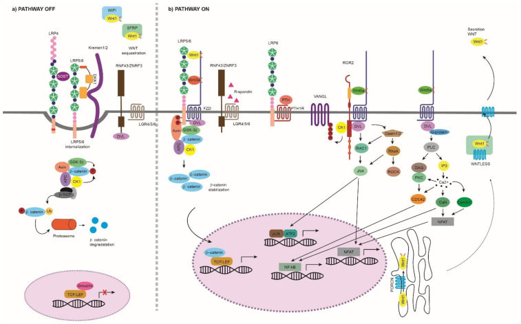
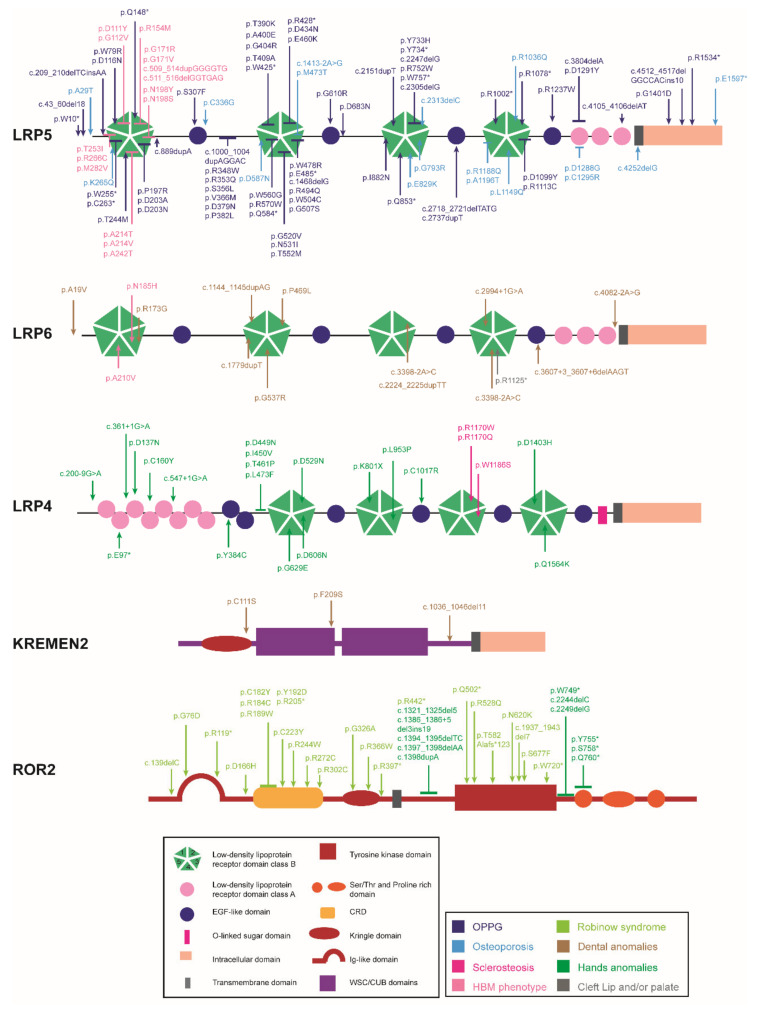
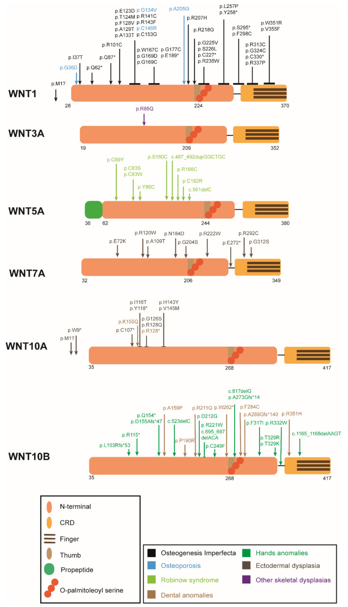
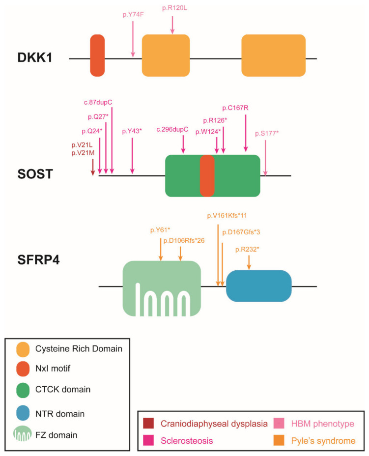
The Wnt pathway is involved in several processes essential for bone development and homeostasis. For proper functioning, the Wnt pathway is tightly regulated by numerous extracellular elements that act by both activating and inhibiting the pathway at different moments. This review aims to describe, summarize and update the findings regarding the extracellular modulators of the Wnt pathway, including co-receptors, ligands and inhibitors, in relation to bone homeostasis, with an emphasis on the animal models generated, the diseases associated with each gene and the bone processes in which each member is involved. The precise knowledge of all these elements will help us to identify possible targets that can be used as a therapeutic target for the treatment of bone diseases such as osteoporosis.

Keywords: Wnt pathway; bone; co-receptors; inhibitors; ligands.


Conflict of interest statement

The authors declare no conflict of interest.

Figures

Figure 1
Figure 1
Wnt pathway. (a) Wnt pathway OFF: The Wnt pathway is inactive in the absence of Wnt ligands or by the effect of extracellular inhibitors that prevent the activation of the pathway. We can group the extracellular inhibitors into two categories depending on the action they perform: (i) binding to the Wnt ligands and preventing their binding to the membrane receptors, such as SFRP1–5 and WIF-1, and (ii) interfering with the LRP binding to FZD, such as DKK1 and sclerostin. Due to their mechanism of action, the first type of inhibitors can act on both canonical and non-canonical pathways, while the second type is specific to the canonical Wnt pathway. The inhibition performed by DKK1 and sclerostin is mediated or enhanced by receptors such as Kremen proteins and LRP4 [27,28]. When the canonical pathway is not activated, β-catenin is phosphorylated at three critical residues and sequestered in the cytoplasm by the destruction complex. The destruction complex is formed by scaffold proteins APC and AXIN1; Ser/Thr kinases, CK1α, ε and δ and GSK3β; and transcriptional regulators YAP/TAZ of the Hippo pathway [29]. This phosphorylated β-catenin is polyubiquitinated for its degradation in the proteasome by the complex containing, among others, β-TrCP protein. Thus, it is not available in the nucleus and, in its absence, a complex is formed by TCF/LEF and TLE/Groucho, inhibiting the transcription of target genes. (b) Wnt pathway ON: The binding of a Wnt ligand to FZD/LRP or ROR1/2/RYK/FZD activates the Wnt pathway that will result in a different transcriptional regulation. Canonical Wnt Pathway ON: The pathway begins at the cell surface with the formation of a heterotrimeric complex consisting of a Wnt ligand (19 different Wnts), a LRP transmembrane co-receptor (LRP5/6) and an FZD receptor (10 different FZDs). The formation of the LRP-FZD-Wnt complex results in phosphorylation of the LRP5/6 co-receptor by CK1α and GSK3β. Then, the DVL (also called DSH) polymerizes and is activated, inhibiting the destruction complex. This produces stabilization and accumulation in the cytoplasm of β-catenin, which will translocate to the nucleus. Once there, it displaces the TLE/Groucho repressors, forming an active complex with TCF/LEF proteins, which results in the recruitment of coactivators and activation of transcription of genes important for the differentiation and formation of bone, such as WISP1 and RUNX2 [12,29]. Non-canonical Wnt Pathway ON: (i) WNT/PCP: The WNT-PCP pathway begins with the binding of a Wnt ligand, such as WNT5A, to FZD and the co-receptors ROR1/2 or RYK. Then, DVL is recruited and activated, resulting in the activation of the scaffold protein VANGL. DVL forms a complex with DAMM1, which activates the small GTPases RHOA and RAC1, which in turn activate ROCK and JNK. This leads to rearrangements of the cytoskeleton and/or the induction of transcription through ATF2 and/or NFAT. The activation of the WNT5A-ROR1/2 pathway inhibits the canonical Wnt signaling [24,25]. (ii) WNT/Ca2+: The binding of the ligand to an FZD receptor results in the recruitment and activation of DVL. Then, DVL binds to the small GTPases, which activate PLC. This activation leads to the breakage of PIP2 into DAG and IP3. When IP3 binds to its receptor on the endoplasmic reticulum, calcium release occurs. When the calcium concentration is increased, DAG activates PKC, which in turn can activate CDC42. Increased intracellular calcium can also activate calcineurin and CaMKII, which in turn can induce the activation of the NFAT transcription factor or NF-kB [26,30]. WNT signaling modulators: The binding of RSPO to LGR and to RNF43/ZNRF3 maintains the Wnt signal ON by preventing the polyubiquitination and endocytosis of FZD performed by RNF43/ZNF3 [31]. WNT production: Wnt precursors undergo post-translational modifications such as porcupine-mediated palmitoylation, other lipid modifications and glycosylation in the ER. Then, the transmembrane protein Wntless (Wls) transports the functional Wnt ligands to the plasma membrane via the golgi apparatus. Wnt ligands are secreted from the cell by solubilization, exosome formation or on lipid protein particles [32,33,34]. See next figures for the information on the different protein domains. APC: adenomatous polyposis coli; AXIN1: axis inhibition protein 1; β-TrCP: β-Transducin repeat-containing protein; CAMKII: calcium/calmodulin-dependent protein kinase II; CDC42: cell division control protein 42; CK1: casein kinase 1; DAAM1: DVL-associated activator of morphogenesis; DVL: disheveled: FZD: frizzled; GSK3β: glycogen synthase kinase 3β; IP3, inositol 1,4,5 triphosphate, JNK: JUN kinase; LGR: leucine-rich repeat-containing G-protein-coupled receptor; NFAT: nuclear factor of activated T cells; NF-kB: nuclear factor kappa B; PKC: protein kinase C; PLC: phospholipase C; RAC: Ras-related C3 botulinum substrate; RHOA: Ras homolog gene family member A; ROCK: Rho kinase; ROR1/2: bind tyrosine kinase-like orphan receptor 1 or 2; RYK: receptor-like tyrosine kinase; PORCN: porcupine; RNF43/ZNRF3: ring finger protein 43/zinc and ring finger 3; RSPO; R-spondin ligand family members; SFRP: secreted frizzled-related proteins; TCF/LEF: T-cell factor/lymphoid enhancer factor; TLE: Transducin-Like Enhancer of Split Proteins; VANGL: Van Gogh-like; YAP/TAZ: Yes-associated protein/transcriptional co-activator with a PDZ-binding domain; WIF-1: Wnt inhibitory factor 1.
Figure 2
Figure 2
Domain structure of the co-receptors and localization of the mutations causing human skeletal diseases according to human gene mutation database (HGMD). ↓ indicates a point mutation, ⊥ encompasses more than one aminoacid position (a cluster of point mutations). *: STOP codon.
Figure 3
Figure 3
Schematic protein representation of the Wnt ligands with the mutations causing human skeletal diseases according to HGMD. ↓ indicates a point mutation, ⊥ encompasses more than one aminoacid position (a cluster of point mutations). Numbers below the structure show the amino acid position in the peptide before post-translational modification. *: STOP codon.
Figure 4
Figure 4
Schematic protein representation of the Wnt inhibitors with the mutations causing human skeletal diseases according HGMD. *: STOP codon.

References

    1. Nusse R., Brown A., Papkoff J., Scambler P., Shackleford G., McMahon A., Moon R., Varmus H. A New Nomenclature for Int-1 and Related Genes: The Wnt Gene Family. Cell. 1991;64:231. doi: 10.1016/0092-8674(91)90633-A. - DOI - PubMed
    1. Nusse R., Varmus H.E. Wnt Genes. Cell. 1992;69:1073–1087. doi: 10.1016/0092-8674(92)90630-U. - DOI - PubMed
    1. Nusse R. Wnt Signaling in Disease and in Development. Cell Res. 2005;15:28–32. doi: 10.1038/sj.cr.7290260. - DOI - PubMed
    1. Van Amerongen R. Alternative Wnt Pathways and Receptors. Cold Spring Harb. Perspect. Biol. 2012;4:a007914. doi: 10.1101/cshperspect.a007914. - DOI - PMC - PubMed
    1. Nalesso G., Sherwood J., Bertrand J., Pap T., Ramachandran M., De Bari C., Pitzalis C., Dell’accio F. WNT-3A Modulates Articular Chondrocyte Phenotype by Activating Both Canonical and Noncanonical Pathways. J. Cell Biol. 2011;193:551–564. doi: 10.1083/jcb.201011051. - DOI - PMC - PubMed

Publication types