

Gut Microbiota-Derived PGF2α Fights against Radiation-Induced Lung Toxicity through the MAPK/NF-κB Pathway
- PMID: 35052569
- PMCID: PMC8773112
- DOI: 10.3390/antiox11010065
Gut Microbiota-Derived PGF2α Fights against Radiation-Induced Lung Toxicity through the MAPK/NF-κB Pathway
Abstract
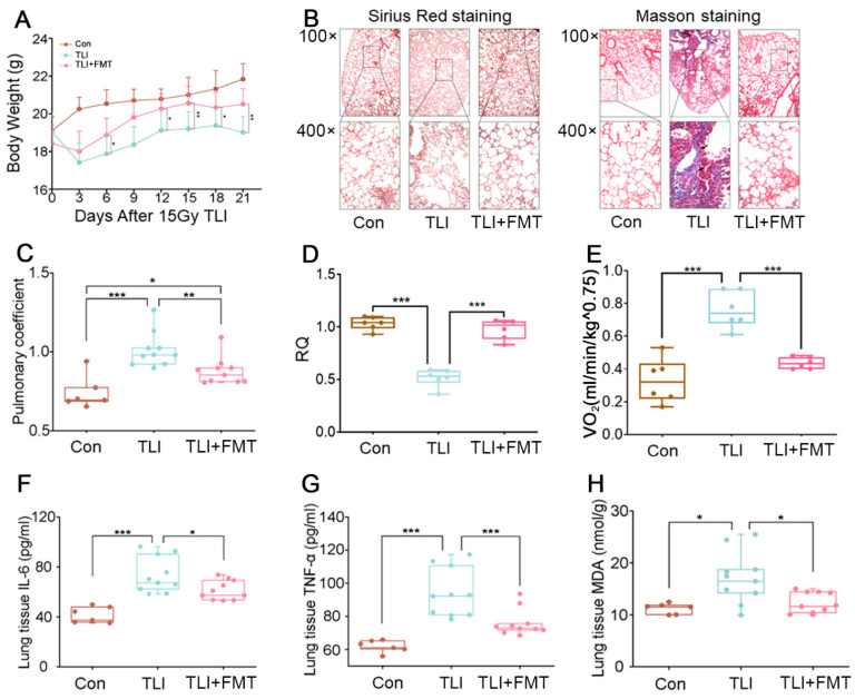
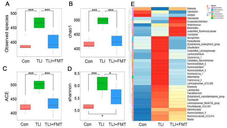
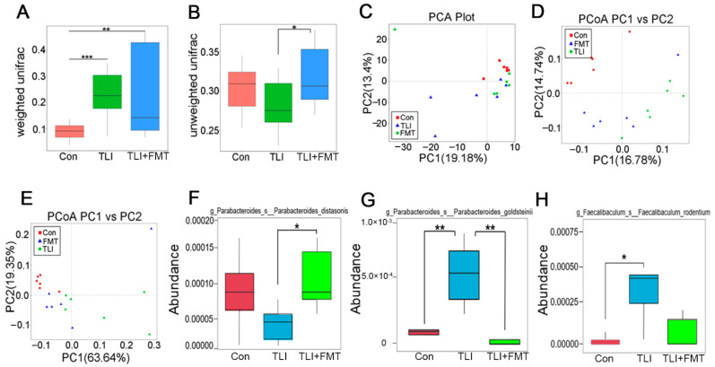
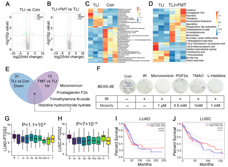
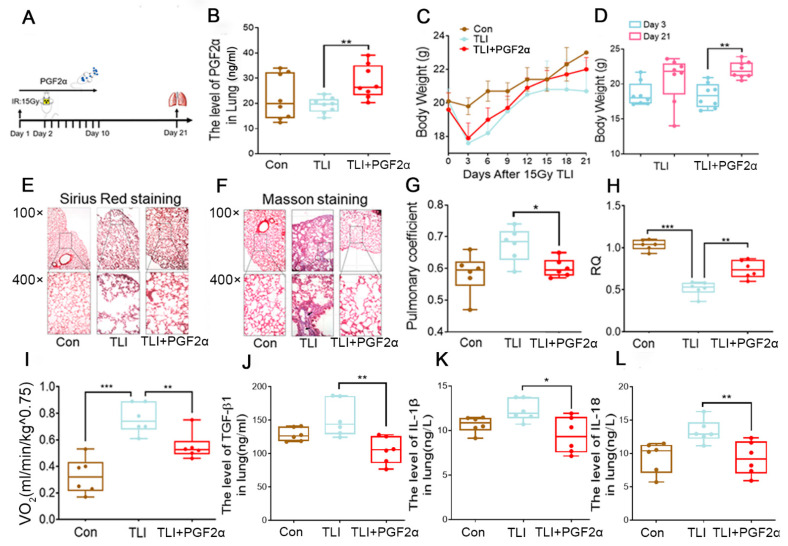
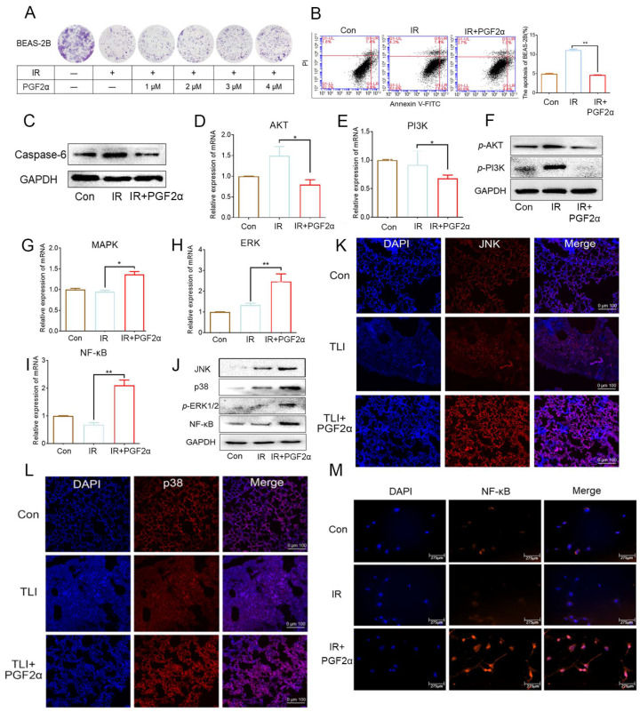
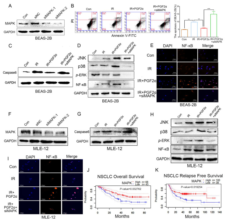
Radiation pneumonia is a common and intractable side effect associated with radiotherapy for chest cancer and involves oxidative stress damage and inflammation, prematurely halting the remedy and reducing the life quality of patients. However, the therapeutic options for the complication have yielded disappointing results in clinical application. Here, we report an effective avenue for fighting against radiation pneumonia. Faecal microbiota transplantation (FMT) reduced radiation pneumonia, scavenged oxidative stress and improved lung function in mouse models. Local chest irradiation shifted the gut bacterial taxonomic proportions, which were preserved by FMT. The level of gut microbiota-derived PGF2α decreased following irradiation but increased after FMT. Experimental mice with PGF2α replenishment, via an oral route, exhibited accumulated PGF2α in faecal pellets, peripheral blood and lung tissues, resulting in the attenuation of inflammatory status of the lung and amelioration of lung respiratory function following local chest irradiation. PGF2α activated the FP/MAPK/NF-κB axis to promote cell proliferation and inhibit apoptosis with radiation challenge; silencing MAPK attenuated the protective effect of PGF2α on radiation-challenged lung cells. Together, our findings pave the way for the clinical treatment of radiotherapy-associated complications and underpin PGF2α as a gut microbiota-produced metabolite.
Keywords: PGF2α; gut microbiota; gut microbiota metabolites; gut-lung axis; radiation pneumonia.
Conflict of interest statement
The authors declare no conflict of interest.
Figures
Similar articles
-
Gut Microbiota-Derived l-Histidine/Imidazole Propionate Axis Fights against the Radiation-Induced Cardiopulmonary Injury.Int J Mol Sci. 2021 Oct 23;22(21):11436. doi: 10.3390/ijms222111436. Int J Mol Sci. 2021. PMID: 34768867 Free PMC article.
-
Gut microbiota-derived indole 3-propionic acid protects against radiation toxicity via retaining acyl-CoA-binding protein.Microbiome. 2020 May 20;8(1):69. doi: 10.1186/s40168-020-00845-6. Microbiome. 2020. PMID: 32434586 Free PMC article.
-
Oral microbiota transplantation fights against head and neck radiotherapy-induced oral mucositis in mice.Comput Struct Biotechnol J. 2021 Oct 25;19:5898-5910. doi: 10.1016/j.csbj.2021.10.028. eCollection 2021. Comput Struct Biotechnol J. 2021. PMID: 34815834 Free PMC article.
-
The Role of Gut Microbiota in Lung Cancer: From Carcinogenesis to Immunotherapy.Front Oncol. 2021 Aug 19;11:720842. doi: 10.3389/fonc.2021.720842. eCollection 2021. Front Oncol. 2021. PMID: 34490119 Free PMC article. Review.
-
Treating From the Inside Out: Relevance of Fecal Microbiota Transplantation to Counteract Gut Damage in GVHD and HIV Infection.Front Med (Lausanne). 2020 Aug 6;7:421. doi: 10.3389/fmed.2020.00421. eCollection 2020. Front Med (Lausanne). 2020. PMID: 32850913 Free PMC article. Review.
Cited by
-
Radiation-induced injury and the gut microbiota: insights from a microbial perspective.Therap Adv Gastroenterol. 2025 Jun 16;18:17562848251347347. doi: 10.1177/17562848251347347. eCollection 2025. Therap Adv Gastroenterol. 2025. PMID: 40535532 Free PMC article. Review.
-
Sexual dimorphism in gut microbiota dictates therapeutic efficacy of intravenous immunoglobulin on radiotherapy complications.J Adv Res. 2023 Apr;46:123-133. doi: 10.1016/j.jare.2022.06.002. Epub 2022 Jun 11. J Adv Res. 2023. PMID: 35700918 Free PMC article.
-
Microbiome in radiotherapy: an emerging approach to enhance treatment efficacy and reduce tissue injury.Mol Med. 2024 Jul 19;30(1):105. doi: 10.1186/s10020-024-00873-0. Mol Med. 2024. PMID: 39030525 Free PMC article. Review.
-
Prostaglandin F2α Regulates Adipogenesis by Modulating Extracellular Signal-Regulated Kinase Signaling in Graves' Ophthalmopathy.Int J Mol Sci. 2023 Apr 10;24(8):7012. doi: 10.3390/ijms24087012. Int J Mol Sci. 2023. PMID: 37108173 Free PMC article.
-
The Effect of Metformin on Radiation-Induced Lung Fibrosis in Mice.Dose Response. 2024 Dec 10;22(4):15593258241308051. doi: 10.1177/15593258241308051. eCollection 2024 Oct-Dec. Dose Response. 2024. PMID: 39664837 Free PMC article.
References
-
- Brainard J., Farver C. The diagnosis of non-small cell lung cancer in the molecular era. Mod. Pathol. 2019;32((Suppl. 1)):16–26. - PubMed
-
- Kasmann L., Dietrich A., Staab-Weijnitz C.A., Manapov F., Behr J., Rimner A., Jeremic B., Senan S., De Ruysscher D., Lauber K., et al. Radiation-induced lung toxicity-cellular and molecular mechanisms of pathogenesis, management, and literature review. Radiat. Oncol. 2020;15:214. doi: 10.1186/s13014-020-01654-9. - DOI - PMC - PubMed
Grants and funding
LinkOut - more resources
Full Text Sources
