. 2021 Dec 21;10(1):2.
doi: 10.3390/biomedicines10010002.

Collagenase-Induced Patellar Tendinopathy with Neovascularization: First Results towards a Piglet Model of Musculoskeletal Embolization

Affiliations

Collagenase-Induced Patellar Tendinopathy with Neovascularization: First Results towards a Piglet Model of Musculoskeletal Embolization

Julien Ghelfi et al. Biomedicines. .

Abstract

Background: Therapeutic strategies targeting neovessels responsible for musculoskeletal chronic pain have emerged, including neovessels embolization. Our study aimed to develop a large animal model of patellar tendinopathy with neovascularization.

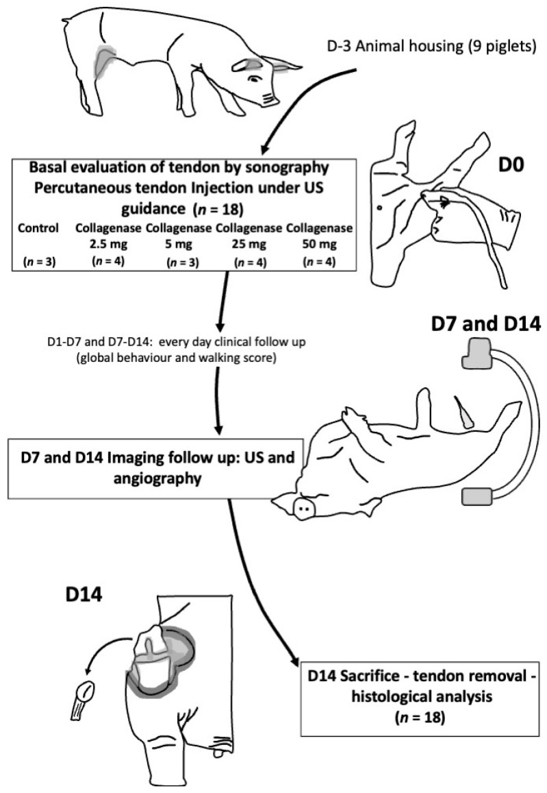
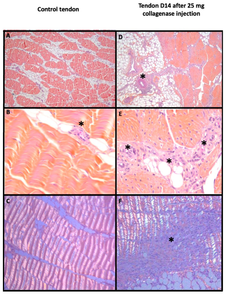
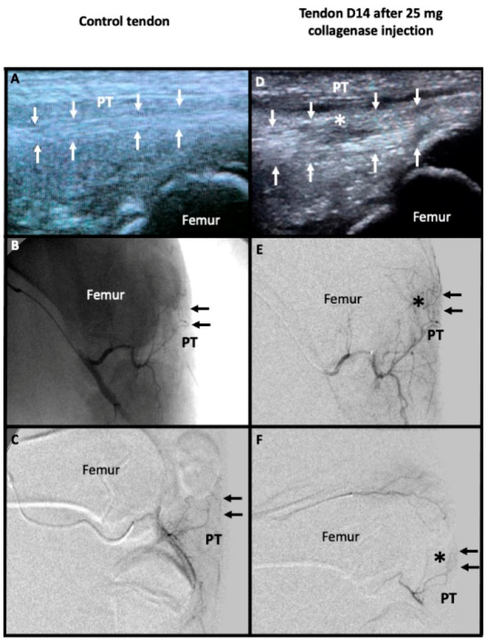
Methods: Nine 3-month-old male piglets (18 patellar tendons) received percutaneous injections of increasing doses of collagenase (0 to 50 mg) at day 0 (D0). Tendinopathy was evaluated by ultrasound (D7 and D14). Neovascularization was evaluated visually and on angiographies. Bonar score was used for histological analysis (D14). Correlations were evaluated using Spearman's rank (Rs) test.

Results: Research protocol was well tolerated. All tendons were enlarged with a median increase of 31.58% [25-40.28] at D7 (p = 0.244) at D7 and 57.52% [48.41-91.45] at D14 (p = 0.065). Tendons with collagenase injection had more hypoechoic changes, with one tendon rupture (p = 0.012). Neovascularization was reported above 5 mg collagenase (p < 0.01) at D7 and D14 with dose-related neovessels induction (Rs = 0.8, p < 0.001). The Bonar score increased above 5 mg collagenase, correlated with the dose (Rs = 0.666, p = 0.003).

Conclusions: The study shows the feasibility, safety and reproducibility of this new large animal model of patellar tendinopathy with neovascularization after collagenase injection. It will allow studying new treatments on direct embolization of neovessels by endovascular approach.

Keywords: animal model; embolization; neovascularization; patellar tendon; tendinopathy.


Conflict of interest statement

The authors declare no conflict of interest.

Figures

Figure 1
Figure 1
Flowchart of the study. US: ultrasound; D: day.
Figure 2
Figure 2
Histological evaluation of a control tendon (left) and a pathological tendon 14 days after injection of 25 mg of collagenase (right). Patellar tendon with HES stain ×50 (A,D), ×400 (B,E) magnification and ×100 with polarization (C,F). In the control tendon, (A,B) show regular bundles of small spindle fibroblasts in wavy collagen with few small capillaries between bundles (black asterisk); (C) depicted the regular and wavy aspect in polarization. In the pathological patellar tendon, fibrinoid necrosis appeared in peri-tendinous artery (black asterisk) in (E) with loss of demarcation of fibers bundles, plump fibroblasts and numerous clusters of capillaries (black asterisk) in (D). (F) showed focal distortion of wavy appearance in polarization (black asterisk).
Figure 3
Figure 3
Imaging evaluation between a control tendon (left) and a pathological tendon 14 days after injection of 25 mg of collagenase (right). Sagittal ultrasound acquisition show the regular hyperechoic fibers of the control patellar tendon (PT) framed by small white arrows in (A). Microcatheter was placed inside the main genicular artery to perform unsubstracted angiography (without bone substraction which is still visible) in (B) and digital substraction angiography showing only the vessels like in (C). PT was marked with small black arrows (B,C,E,F). There were no abnormal microvessels in projection of PT in the control tendon (B,C). The pathological tendon showed a marked thickening framed by small white arrows with hypoechoic change in its mid portion (white asterisk) in (D). Angiography in the main genicular artery (E) and in the superior genicular artery (F) showed abnormal microvessels in projection of PT (black asterisk).

References

    1. Dominick K.L., Ahern F.M., Gold C.H., Heller D.A. Health-Related Quality of Life and Health Service Use among Older Adults with Osteoarthritis. Arthritis Care Res. 2004;51:326–331. doi: 10.1002/art.20390. - DOI - PubMed
    1. Neogi T. The Epidemiology and Impact of Pain in Osteoarthritis. Osteoarthr. Cartil. 2013;21:1145–1153. doi: 10.1016/j.joca.2013.03.018. - DOI - PMC - PubMed
    1. Brockmeyer M., Diehl N., Schmitt C., Kohn D.M., Lorbach O. Results of Surgical Treatment of Chronic Patellar Tendinosis (Jumper’s Knee): A Systematic Review of the Literature. Arthroscopy. 2015;31:2424–2429.e3. doi: 10.1016/j.arthro.2015.06.010. - DOI - PubMed
    1. Kettunen J.A., Kvist M., Alanen E., Kujala U.M. Long-Term Prognosis for Jumper’s Knee in Male Athletes. A Prospective Follow-up Study. Am. J. Sports Med. 2002;30:689–692. doi: 10.1177/03635465020300051001. - DOI - PubMed
    1. Millar N.L., Silbernagel K.G., Thorborg K., Kirwan P.D., Galatz L.M., Abrams G.D., Murrell G.A.C., McInnes I.B., Rodeo S.A. Tendinopathy. Nat. Rev. Dis. Primers. 2021;7:1. doi: 10.1038/s41572-020-00234-1. - DOI - PubMed

Grants and funding

LinkOut - more resources