. 2022 Jan 17;10(1):187.
doi: 10.3390/biomedicines10010187.

Effect of Ulinastatin on Syndecan-2-Mediated Vascular Damage in IDH2-Deficient Endothelial Cells

Affiliations

Effect of Ulinastatin on Syndecan-2-Mediated Vascular Damage in IDH2-Deficient Endothelial Cells

Su-Jeong Choi et al. Biomedicines. .

Abstract

Syndecan-2 (SDC2), a cell-surface heparin sulfate proteoglycan of the glycocalyx, is mainly expressed in endothelial cells. Although oxidative stress and inflammatory mediators have been shown to mediate dysfunction of the glycocalyx, little is known about their role in vascular endothelial cells. In this study, we aimed to identify the mechanism that regulates SDC2 expression in isocitrate dehydrogenase 2 (IDH2)-deficient endothelial cells, and to investigate the effect of ulinastatin (UTI) on this mechanism. We showed that knockdown of IDH2 induced SDC2 expression in human umbilical vein endothelial cells (HUVECs). Matrix metalloproteinase 7 (MMP7) influences SDC2 expression. When IDH2 was downregulated, MMP7 expression was increased, as was TGF-β signaling, which regulates MMP7. Inhibition of MMP7 activity using MMP inhibitor II significantly reduced SDC2, suggesting that IDH2 mediated SDC2 expression via MMP7. Moreover, expression of SDC2 and MMP7, as well as TGF-β signaling, increased in response to IDH2 deficiency, and treatment with UTI reversed this increase. Similarly, the increase in SDC2, MMP7, and TGF-β signaling in the aorta of IDH2 knockout mice was reversed by UTI treatment. These findings suggest that IDH2 deficiency induces SDC2 expression via TGF-β and MMP7 signaling in endothelial cells.

Keywords: IDH2; SDC2; endothelial cells; ulinastatin; vascular damage.


Conflict of interest statement

The authors declare no conflict of interest.

Figures

Figure 1
Figure 1
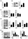
Expression of Syndecans in IDH2 deficient HUVECs. HUVECs were transfected with dose dependent siIDH2 (10 pmol, 50 pmol) and siCON for 48h. mRNA expressions of (a) SDC1, (b) SDC2, (c) SDC3, (d) SDC4 were quantified by qPCR (e) protein level of SDC2 was detected by western blotting. β-actin was used as a loading control. Protein levels were quantified by densitometric analysis using Image-J software (Shown in the right panel). (f) Immunofluorescence analysis of HUVECs to compare the SDC2 expression in siCON and siIDH2 (50 pmol) transfected HUVECs (scale bar 100 μm). All data are presented as means ± SEM of three independent experiments, * p < 0.05, compared with siCON.
Figure 2
Figure 2
Involvement of TGF-β and MMP7 signaling in relation to SDC2 shedding in IDH2 deficient HUVECs. HUVECs were transfected with 50 pmol of siIDH2 and siCON for 48 h. (a) mRNA expression of TGF-β was quantified by qPCR. Protein levels of (b) MMP7 (c) β-catenin and (d) TGF-β signaling-related proteins were detected by western blotting. β-actin was used as a loading control. Protein levels were quantified by densitometric analysis using Image-J software (Shown in the right panel). HUVECs were transfected with 50 pmol of siIDH2 and siCON, incubated for 24 h and treated with MMP inhibitor II (25 μM) for another 24 h. Protein levels of (e) MMP7 and SDC2 were detected by western blotting. β-actin was used as a loading control. Protein levels were quantified by densitometric analysis using Image-J software (Shown in the right panel). All data are presented as means ± SEM of three independent experiments, * p < 0.05, compared with siCON, # p < 0.05 compared with the 50 pmol siIDH2 treated cells.
Figure 3
Figure 3
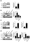
Effect of UTI on SDC2, MMP7 and TGF-β signaling in IDH2 deficient HUVECs. HUVECs were transfected with 50 pmol of siIDH2 and siCON and 1000 U/mL of UTI. mRNA expressions of (a) SDC2 and (c) MMP7 were quantified by qPCR. Protein levels of (b) SDC2 and (d) MMP7 were detected by western blotting. (e) Protein level of TGF-β signaling was detected by western blotting. β-actin was used as a loading control. Protein levels were quantified by densitometric analysis using Image-J software (Shown in the right panel). All data are presented as means ± SEM of three independent experiments, * p < 0.05, compared with siCON, # p < 0.05 compared with the 50 pmol siIDH2 treated cells.
Figure 4
Figure 4
Effect of UTI on SDC2, MMP7 and TGF-β signaling in aorta from IDH2 KO mice. Aorta were isolated from WT and IDH2 KO mice and after peritoneal injection of UTI in WT and IDH2 KO mice. (a,b) Protein level of SDC2, (c,d) TGF-β and MMP7 were detected by western blotting. β-actin was used as a loading control. Protein levels were quantified by densitometric analysis using Image-J software (Shown in the right panel). All data are presented as means ± SEM, * p < 0.05 compared with WT mice, # p < 0.05 compared with UTI injected IDH2 KO mice (n = 10 mice/group).
Figure 5
Figure 5
Schematic representation of the proposed pathway of SDC2 activation in IDH2 deficiency condition.

References

    1. Weinbaum S., Tarbell J.M., Damiano E.R. The structure and function of the endothelial glycocalyx layer. Annu. Rev. Biomed. Eng. 2007;9:121–167. doi: 10.1146/annurev.bioeng.9.060906.151959. - DOI - PubMed
    1. Tarbell J.M., Simon S.I., Curry F.-R.E. Mechanosensing at the vascular interface. Annu. Rev. Biomed. Eng. 2014;16:505–532. doi: 10.1146/annurev-bioeng-071813-104908. - DOI - PMC - PubMed
    1. Fu B.M., Tarbell J.M. Mechano-sensing and transduction by endothelial surface glycocalyx: Composition, structure, and function. WIREs Syst. Biol. Med. 2013;5:381–390. doi: 10.1002/wsbm.1211. - DOI - PMC - PubMed
    1. Tarbell J.M., Shi Z.-D., Dunn J., Jo J. Fluid mechanics, arterial disease, and gene expression. Annu. Rev. Fluid Mech. 2014;46:591–614. doi: 10.1146/annurev-fluid-010313-141309. - DOI - PMC - PubMed
    1. Becker B.F., Jacob M., Leipert S., Salmon A.H.J., Chappell D. Degradation of the endothelial glycocalyx in clinical settings: Searching for the sheddases. Br. J. Clin. Pharmacol. 2015;80:389–402. doi: 10.1111/bcp.12629. - DOI - PMC - PubMed

LinkOut - more resources