

Effect of Displacement Degree of Distal Chevron Osteotomy on Metatarsal Stress: A Finite Element Method
- PMID: 35053125
- PMCID: PMC8772834
- DOI: 10.3390/biology11010127
Effect of Displacement Degree of Distal Chevron Osteotomy on Metatarsal Stress: A Finite Element Method
Abstract
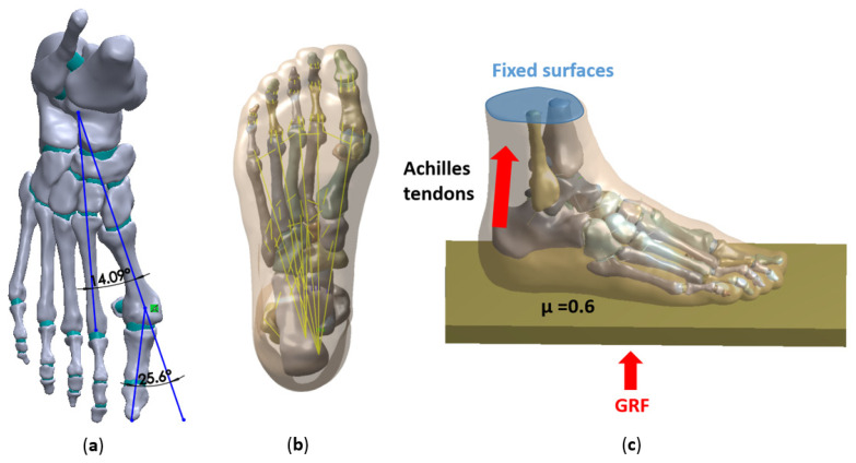
The stress of foot bone can effectively evaluate the functional damage caused by foot deformity and the results of operation. In this study, the finite element method was used to investigate the degree of displacement of distal chevron osteotomy on metatarsal stress and metatarsophalangeal joint load; Methods: Four finite element models of displacement were established by using the CT images of a patient with moderate hallux valgus (hallux valgus angle and intermetatarsal angle were 26.74° and 14.09°, respectively), and the validity of the model was verified. Each finite element model consisted of bones and various cartilage structures, ligaments, and plantar fascia, as well as encapsulated soft tissue. Except for soft tissue, the material properties of other parts were isotropic linear elastic material, and the encapsulated soft tissue was set as nonlinear hyperelastic material. The mesh was tetrahedral mesh. Link elements were used in ligament and plantar fascia. A ground reaction force with a half-body weight was applied at the bottom of the floor to simulate the ground reaction when standing. The upper surfaces of the encapsulated soft tissue, distal tibia, and distal fibula were fixed. The stress distribution of metatarsals and the stress of cartilage of the first metatarsophalangeal joint were compared and analyzed; Results: Compared with the hallux valgus without osteotomy, the stress of the first metatarsals and second metatarsals of 2-4 mm decreased, and the stress of the interarticular cartilage of the first metatarsophalangeal joint with 4 mm was reduced. In the case of 6 mm, the stress value between the first metatarsal and the first metatarsophalangeal joint increased, and 4 mm was the most suitable distance; Conclusions: Compared with the hallux valgus without osteotomy, the stress of the first metatarsals and second metatarsals of 2-4 mm decreased, and the stress of the interarticular cartilage of the first metatarsophalangeal joint with 4 mm was reduced. In the case of 6 mm, the stress value between the first metatarsal and the first metatarsophalangeal joint increased, and 4 mm was the most suitable distance. For the degree of displacement of the distal chevron osteotomy, the postoperative stability and the stress distribution of metatarsal bone should be considered. Factors such as hallux valgus angle, intermetatarsal angle, patient's age, body weight, and metatarsal width should be considered comprehensively. The factors affecting osteotomy need to be further explored. The degree of displacement of osteotomy can be evaluated by FE method before the operation, and the most suitable distance can be obtained.
Keywords: chevron osteotomy; hallux valgus; metatarsal stress; metatarsophalangeal stress.
Conflict of interest statement
The authors declare no conflict of interest.
Figures


References
-
- Zhang B., Lu Q. A Current Review of Foot Disorder and Plantar Pressure Alternation in the Elderly. Phys. Act. Health. 2020;4:95–106. doi: 10.5334/paah.57. - DOI
Grants and funding
LinkOut - more resources
Full Text Sources
