. 2021 Dec 22;12(1):15.
doi: 10.3390/biom12010015.

Connexin 43 Gene Ablation Does Not Alter Human Pluripotent Stem Cell Germ Lineage Specification

Affiliations

Connexin 43 Gene Ablation Does Not Alter Human Pluripotent Stem Cell Germ Lineage Specification

Grace A Christopher et al. Biomolecules. .

Abstract

During embryonic germ layer development, cells communicate with each other and their environment to ensure proper lineage specification and tissue development. Connexin (Cx) proteins facilitate direct cell-cell communication through gap junction channels. While previous reports suggest that gap junctional intercellular communication may contribute to germ layer formation, there have been limited comprehensive expression analyses or genetic ablation studies on Cxs during human pluripotent stem cell (PSC) germ lineage specification. We screened the mRNA profile and protein expression patterns of select human Cx isoforms in undifferentiated human induced pluripotent stem cells (iPSCs), and after directed differentiation into the three embryonic germ lineages: ectoderm, definitive endoderm, and mesoderm. Transcript analyses by qPCR revealed upregulation of Cx45 and Cx62 in iPSC-derived ectoderm; Cx45 in mesoderm; and Cx30.3, Cx31, Cx32, Cx36, Cx37, and Cx40 in endoderm relative to control human iPSCs. Generated Cx43 (GJA1) CRISPR-Cas9 knockout iPSCs successfully differentiated into cells of all three germ layers, suggesting that Cx43 is dispensable during directed iPSC lineage specification. Furthermore, qPCR screening of select Cx transcripts in our GJA1-/- iPSCs showed no significant Cx upregulation in response to the loss of Cx43 protein. Future studies will reveal possible compensation by additional Cxs, suggesting targets for future CRISPR-Cas9 ablation studies in human iPSC lineage specification.

Keywords: CRISPR-Cas9 gene ablation; connexin channels; differentiation; germ lineage specification; human pluripotent stem cells.


Conflict of interest statement

The authors declare no conflict of interest.

Figures
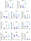
Figure 1
Figure 1
Connexin mRNA expression during human iPSC lineage specification. Quantitative RT-PCR (qPCR) expression of mRNA transcripts encoding 11 of the 21 human connexin isoforms in human control (CTL) iPSCs (grey dotted line) and after directed differentiation into ectoderm, endoderm, and mesoderm. Data represent the standard error of the mean of 3–5 independent experiments. * p < 0.05; ** p < 0.01 compared to undifferentiated iPSCs.
Figure 2
Figure 2
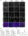
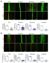
Cx43 protein expression and localization in iPSCs and cells of each germ lineage. (A) Representative immunofluorescent confocal micrographs, demonstrating Cx43 (green) localization in control iPSCs (SOX2, iPSCs: purple) and after directed differentiation toward the three germ lineages (Nestin, ectoderm; Brachyury, mesoderm; SOX17, endoderm: purple) and nuclei (Hoechst, nuclei: blue). Cx43 forms large puncta (white arrows) at the cell surface indicative of gap junction plaques. Scale bars = 50 µm. (B) Representative Western blots and (C) densitometric analysis of total Cx43 protein expression in PAX6-positive ectoderm, Brachyury-positive mesoderm, and SOX17-positive endoderm cells. * p < 0.05 compared to undifferentiated iPSCs. Data represent the standard error of the mean of 3–4 independent experiments.
Figure 3
Figure 3
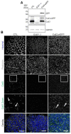
CRISPR-Cas9 manipulation of Cx43 (GJA1) in human iPSCs. (A) Representative Western blots of green fluorescence protein (GFP), Cx43, and GAPDH in control, Cx43 knockout (GJA1-/-), and Cx43eGFP iPSCs. (B) Immunofluorescence confocal microscopy of Cx43 (green) in control, GJA1-/-, and Cx43-eGFP iPSCs. Actin (phalloidin, grey); nuclei (Hoechst, blue). Cx43 forms large puncta (white arrows) at the cell surface indicative of gap junction plaques. Scale bars = 50 µm.
Figure 4
Figure 4
Control and Cx43-eGFP iPSCs spontaneously differentiate into cells from all three germ lineages. Representative immunofluorescence confocal micrographs demonstrating Cx43 or Cx43-eGFP (green) localization in control (A) or Cx43-eGFP reporter (B) iPSCs that underwent spontaneous differentiation into cells of each germ lineage: ectoderm (Nestin, purple), mesoderm (Brachyury, purple), or endoderm (SOX17, purple). Nuclei (Hoechst, blue). Cx43 forms large puncta (white arrows) at the cell surface indicative of gap junction plaques. Scale bars = 50 µm.
Figure 5
Figure 5
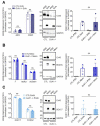
Cx43 knockout iPSCs successfully differentiate to cells of each germ lineage under directed differentiation conditions. Quantitative RT-PCR (qPCR) screen, representative Western blots, and densitometry of differentiated control and Cx43 knockout (GJA1-/-) iPSCs. (A) Ectoderm formation indicated by presence of NES and PAX6 transcripts and expression of PAX6 protein. (B) Mesoderm evaluated by identifying transcripts for T, MILX1, and NCAM1, and expression of Brachyury protein. (C) Endoderm formation confirmed by FOXA2 and SOX17 transcripts and expression of SOX17 protein. Data represent the standard error of the mean of 3–4 independent experiments. ns: not significant compared to control cells.
Figure 6
Figure 6
Spontaneously differentiated Cx43 knockout iPSCs form cells of all three germ lineages. (A) Phase contrast images of control and GJA1-/- embryoid bodies, scale bar 500 µm. (B) qPCR and (C) flow cytometric analyses demonstrate comparable spontaneous differentiation capacities of germ lineage populations by control and GJA1-/- embryoid bodies (pluripotency, grey; ectoderm, dark blue; mesoderm, blue; endoderm, light blue). ns: not significant compared to control cells.
Figure 7
Figure 7
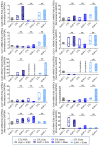
Other connexin isoforms do not compensate for the loss of Cx43 during iPSC lineage commitment. Quantitative RT-PCR (qPCR) screen of mRNA transcripts encoding 10 of the 21 human connexin isoforms in ectoderm, mesoderm, or endoderm directed differentiated from control and GJA1-/- iPSCs. Data represent the standard error of the mean of 4 independent experiments. ns: not significant compared to undifferentiated control iPSCs.
Figure 8
Figure 8
Gap junction coupling is lost in Cx43 knockout iPSCs and cells of each germ lineage. Scrape loading dye transfer assay of control and GJA1-/- iPSCs, ectoderm, mesoderm, and endoderm cells. (A) Representative fluorescence images of gap junction permeable dye Lucifer yellow (LY) or (B) neurobiotin. Scale bars = 200 µm. Data represent the standard error of the mean of 3–5 independent experiments. * p < 0.05; ** p < 0.01, *** p < 0.001 compared to control cells.
Figure 9
Figure 9
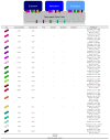
Human connexin expression profiles described in literature and this study. Human plu-ripotent stem cells express Cx40.1 [21] and Cx50 [21]. Cx23 expression in pluripotent stem cells and the three germ layers has not yet been characterized. Our study shows control iPSCs and the three germ layers express mRNA transcripts for Cx26 [21,38,51], Cx30.3 [7,21,38], Cx31 [7,21,38], Cx31.1 [7,21,38], Cx37 [7,21,38], Cx40 [7,21,38], Cx43 [7,21,22,27,38,43,44,45], Cx45 [7,21,38,45], and Cx62 [7,21,38]. Significant increases were viewed for mRNA transcripts of Cx62 in ectoderm; Cx45 in ectoderm and mesoderm; and for Cx30.3, Cx31, Cx32, Cx36, Cx37, and Cx40 in endo-derm. Transcripts for Cx32 and Cx36 were not detected in mesoderm.

References

    1. Wörsdörfer P., Maxeiner S., Markopoulos C., Kirfel G., Wulf V., Auth T., Urschel S., von Maltzahn J., Willecke K. Connexin Expression and Functional Analysis of Gap Junctional Communication in Mouse Embryonic Stem Cells. Stem Cells. 2008;26:431–439. doi: 10.1634/stemcells.2007-0482. - DOI - PubMed
    1. Esseltine J.L., Laird D.W. Next-Generation Connexin and Pannexin Cell Biology. Trends Cell Biol. 2016;26:944–955. doi: 10.1016/j.tcb.2016.06.003. - DOI - PubMed
    1. Laird D.W. Life Cycle of Connexins in Health and Disease. Biochem. J. 2006;394:527–543. doi: 10.1042/BJ20051922. - DOI - PMC - PubMed
    1. Bevans C.G., Kordel M., Rhee S.K., Harris A. Isoform Composition of Connexin Channels Determines Selectivity among Second Messengers and Uncharged Molecules. J. Biol. Chem. 1998;273:2808–2816. doi: 10.1074/jbc.273.5.2808. - DOI - PubMed
    1. Harris A.L., Contreras J.E. Motifs in the Permeation Pathway of Connexin Channels Mediate Voltage and Ca2+ Sensing. Front. Physiol. 2014;5:113. doi: 10.3389/fphys.2014.00113. - DOI - PMC - PubMed

Publication types

LinkOut - more resources