. 2022 Jan 13;11(2):265.
doi: 10.3390/cells11020265.

Revisiting the Non-Coding Nature of Pospiviroids

Affiliations

Revisiting the Non-Coding Nature of Pospiviroids

Konstantina Katsarou et al. Cells. .

Abstract

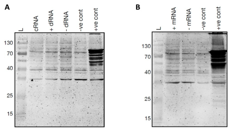
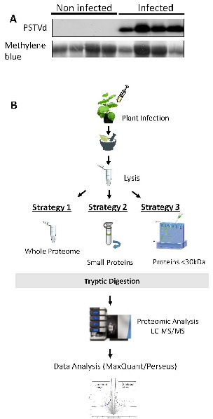
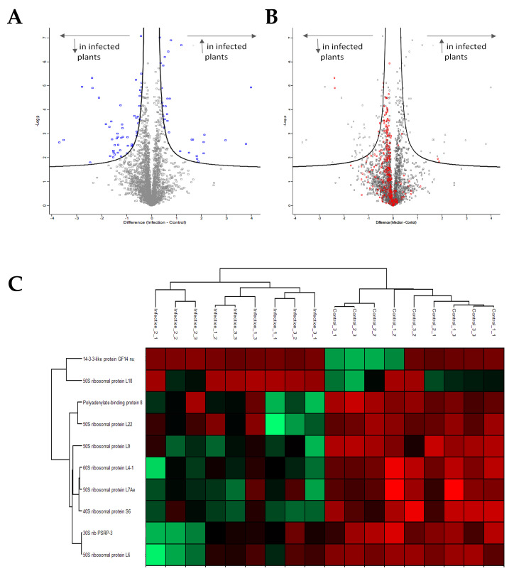
Viroids are small, circular, highly structured pathogens that infect a broad range of plants, causing economic losses. Since their discovery in the 1970s, they have been considered as non-coding pathogens. In the last few years, the discovery of other RNA entities, similar in terms of size and structure, that were shown to be translated (e.g., cirRNAs, precursors of miRNA, RNA satellites) as well as studies showing that some viroids are located in ribosomes, have reignited the idea that viroids may be translated. In this study, we used advanced bioinformatic analysis, in vitro experiments and LC-MS/MS to search for small viroid peptides of the PSTVd. Our results suggest that in our experimental conditions, even though the circular form of PSTVd is found in ribosomes, no produced peptides were identified. This indicates that the presence of PSTVd in ribosomes is most probably not related to peptide production but rather to another unknown function that requires further study.

Keywords: PSTVd; cirRNA; mass spectrometry; non-coding RNA; translation; viroid.


Conflict of interest statement

The authors declare that there is no conflict of interest. This work does not contain any human/animal experiments, and no personal information was used.

Figures

Figure 1
Figure 1
Identification of Possible ORFs in PSTVd. (A) Conservation rate in PSTVd isolates. (B) Comparison between artificially shuffled genome and real genome for PSTVd. (C) Presence of ‘hotspots’ in PSTVd genome.
Figure 2
Figure 2
Identification of possible quasi-species using viroid-derived siRNA and total RNA NGS analysis. (A,C) To locate the potential translation start codons on the PSTVdRG1 and PSTVdNB molecule, the in silico detected alternate start codons (indicated by green line over the nucleotides), the point mutation that could lead into a start codon (blue font), and the stop codons (red font) are shown on secondary structure of PSTVd. The green letters indicate the different nucleotides between PSTVdRG1 and PSTVdNB. (B) Analysis of sRNA derived from PSTVdRG1-inoculated plants revealed the presence of translation start codon (AUG) on PSTVdRG1 sequence. Location and changes in sequence variation that lead into the formation of potential start codons are shown on the secondary structure of PSTVdRG1. The red font indicates the nucleotide that was changed during infection. The two or three mutations that led into the formation of AUG are shown by blue font and an asterisk (*) indicates the nucleotide that showed both point mutation and double mutation. (D) Colors represent the same as in B but for PSTVdNB. However, only the mutations with the higher percentage range per position are represented in this figure (described in Table S4).
Figure 3
Figure 3
Detection of ribosome-associated PSTVd in host plants. Both Tomato cv. Rutgers and N. benthamiana plants were inoculated with PSTVdRG1. (A) Total RNA extracted and RT-PCR assay from these plants at 3 wpi was used to monitor the PSTVd infection. Lane L (Ladder); TC (tomato control), mock inoculated tomato plants; TP, PSTVdRG1 inoculated tomato plants; BC (N. benthamiana control), mock inoculated N. benthamiana plants; BP, PSTVdRG1-inoculated N. benthamiana plants; +ve, RT-PCR positive control; RT ve, RT negative control and, ve, PCR negative control. (B) Flow chart illustrating the details of the isolation of total ribosomes from leaf samples (see Materials and Methods). The resulting precipitates were subjected to RNA purification and analyzed by (C) RT-PCR and (D) Northern blot assays. The lanes were loaded as in (C). (E) RT-qPCR to evaluate the enrichment of PSTVdRG1 in the ribosomes. The expression change is presented on a log2 scale. Error bars indicate the standard deviation (SD).
Figure 4
Figure 4
Polysome fractionation. (A) Flow chart illustrating the details of the separation of the 40S, the 60S and 80S ribosomes and of the polysomes. (B) RNA isolated from the fractionated non-translating ribosomes and from the polysomes were subjected to the RT-PCR assay using the Vid-FW/Vid-RE primer pair. Ladder (L); RNA extracted from mock inoculated tomato plants (TC), PSTVd inoculated tomato plants (TP), mock inoculated N. benthamiana plants (BC) and PSTVd inoculated N. benthamiana plants (BP). RNA extracted from non-translating ribosomes is indicated as NTR, and the RNA extracted from the polysome fraction is denoted by PS. +ve, RT-PCR positive control; RT ve, RT negative control; and ve, PCR negative control. (C) Schematic representation of the differentiation of circular PSTVd RNA by RT-PCR assay. In the figure, the red right arrowhead indicates the Vid-FW primer, red left arrowhead indicates the Vid-RE primer, blue right arrowhead indicates the PSTVd-254F primer, and the blue left arrowhead indicates the PSTVd-253R primer. R indicates the reverse primer and F indicates the forward primer. The black dotted lines indicate the cRNA, the red dotted lines indicate the PCR product obtained with the Vid-FW/Vid-RE primer pair and the blue dotted lines indicates the PCR product obtained with the PSTVd-254F/253R primer pair. Vid-FW is complementary to nucleotide positions 355-16 of PSTVdRG1, Vid-RE is complementary to positions 354-336 of PSTVdRG1, PSTVd-254F is complementary to positions 254-273 of PSTVdRG1 and, PSTVd-253R is complementary to positions 253-234 of PSTVdRG1. The number 1 indicates the first nucleotide of PSTVdRG1, and the number 359 indicates the last nucleotide of PSTVdRG1. (D) PCR performed on the cDNA generated by the Vid-RE primer using the PSTVd-254F/PSTVd-253R primer set. The lanes are loaded as shown for (B).
Figure 5
Figure 5
In vitro translation of PSTVdRG1. In vitro translation of (A) circular RNA (cRNA), dimeric (+) PSTVd RNA (+ dRNA), dimeric (−) PSTVd RNA (-dRNA) and (B) monomeric (+) PSTVd RNA (+ mRNA), monomeric (−) PSTVd RNA (- mRNA). A reaction mixture without any template RNA was used as negative control (ve cont), and luciferase control RNA was used as the positive control (+ve cont).
Figure 6
Figure 6
Experimental design for MS experiments. (A) Northern blot for the detection of PSTVdNB in N. benthamiana plants. Total RNA staining (methylene blue) was used as loading control. (B) Three different strategies were followed in this study. In strategy 1, total lysate from both infected and non-infected plants was used for further MS analysis. In strategy 2, total lysate was filtered through specialized column to keep only small peptides, and then proceed with MS analysis. In strategy 3, a 15% polyacrylamide gel was used to separate proteins and only proteins smaller than 30 kDa were kept for further MS analysis.
Figure 7
Figure 7
MS analysis. (A) Volcano plot showing all proteins affected in PSTVd-infected N. benthamiana plants. In total, 85 proteins were shown to be statistically affected. (B) Volcano plot showing proteins related to the translation mechanism affected upon PSTVd infection of N. benthamiana plants. (C) Heat map of proteins related to translation statistically affected by the infection of PSTVd. All graphs were created using the Perseus 1.6.10.43 software [52]. More details in Table S5.

References

    1. Parsyan A. Translation and Its Regulation in Cancer Biology and Medicine. Springer Publishing; Cham, Switzerland: 2014.
    1. Kearse M.G., Wilusz J.E. Non-AUG translation: A new start for protein synthesis in eukaryotes. Genes Dev. 2017;31:1717–1731. doi: 10.1101/gad.305250.117. - DOI - PMC - PubMed
    1. Wu J., Xiao J., Zhang Z., Wang X., Hu S., Yu J. Ribogenomics: The Science and Knowledge of RNA. Genom. Proteom. Bioinform. 2014;12:57–63. doi: 10.1016/j.gpb.2014.04.002. - DOI - PMC - PubMed
    1. Katsarou K., Rao A., Tsagris M., Kalantidis K. Infectious long non-coding RNAs. Biochime. 2015;117:37–47. doi: 10.1016/j.biochi.2015.05.005. - DOI - PubMed
    1. Hsu M.-T., Coca-Prados M. Electron microscopic evidence for the circular form of RNA in the cytoplasm of eukaryotic cells. Nature. 1979;280:339–340. doi: 10.1038/280339a0. - DOI - PubMed

Publication types