

The Role of Structure MRI in Diagnosing Autism
- PMID: 35054330
- PMCID: PMC8774643
- DOI: 10.3390/diagnostics12010165
The Role of Structure MRI in Diagnosing Autism
Abstract
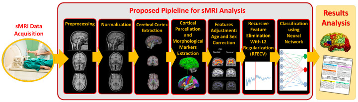
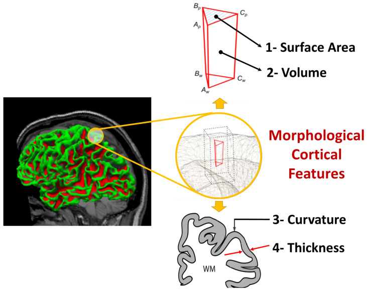
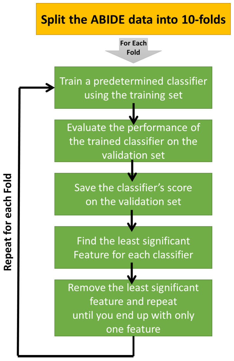
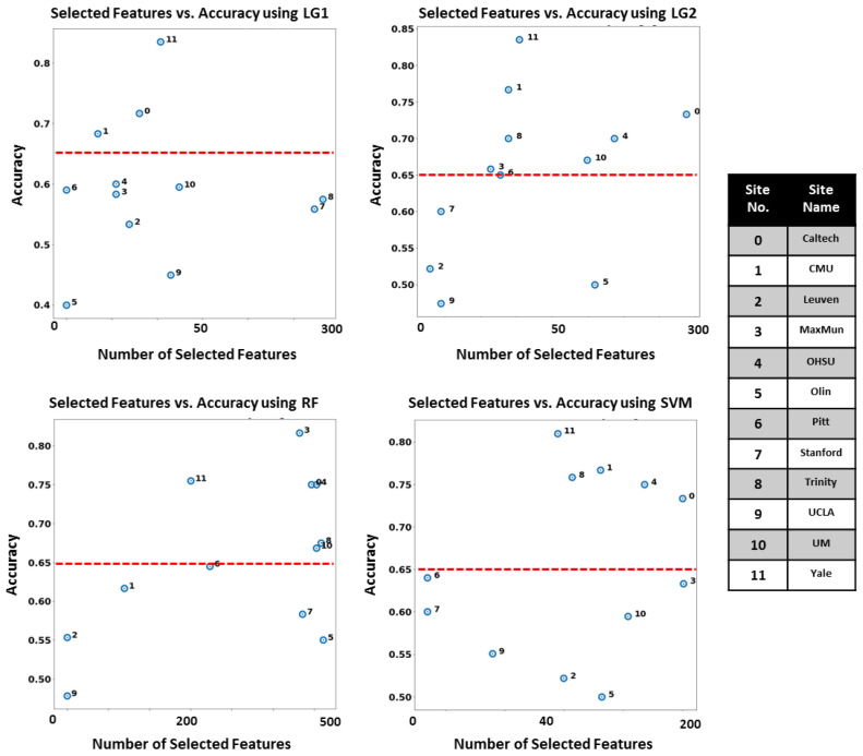
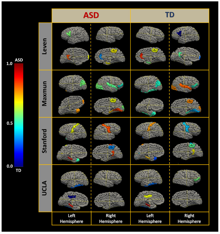
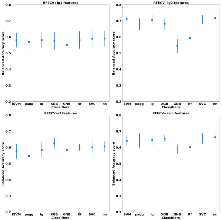
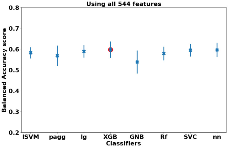
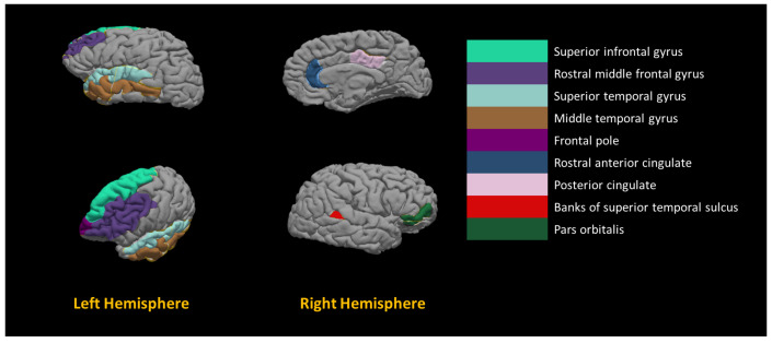
This study proposes a Computer-Aided Diagnostic (CAD) system to diagnose subjects with autism spectrum disorder (ASD). The CAD system identifies morphological anomalies within the brain regions of ASD subjects. Cortical features are scored according to their contribution in diagnosing a subject to be ASD or typically developed (TD) based on a trained machine-learning (ML) model. This approach opens the hope for developing a new CAD system for early personalized diagnosis of ASD. We propose a framework to extract the cerebral cortex from structural MRI as well as identifying the altered areas in the cerebral cortex. This framework consists of the following five main steps: (i) extraction of cerebral cortex from structural MRI; (ii) cortical parcellation to a standard atlas; (iii) identifying ASD associated cortical markers; (iv) adjusting feature values according to sex and age; (v) building tailored neuro-atlases to identify ASD; and (vi) artificial neural networks (NN) are trained to classify ASD. The system is tested on the Autism Brain Imaging Data Exchange (ABIDE I) sites achieving an average balanced accuracy score of 97±2%. This paper demonstrates the ability to develop an objective CAD system using structure MRI and tailored neuro-atlases describing specific developmental patterns of the brain in autism.
Keywords: CAD; autism; classification; feature selection; hyper-parameter optimization; machine learning; structure MRI.
Conflict of interest statement
The authors declare no conflict of interest.
Figures

References
-
- Baio J., Wiggins L., Christensen D.L., Maenner M.J., Daniels J., Warren Z., Kurzius-Spencer M., Zahorodny W., Rosenberg C.R., White T., et al. Prevalence of autism spectrum disorder among children aged 8 years—Autism and developmental disabilities monitoring network, 11 sites, United States, 2014. MMWR Surveill. Summ. 2018;67:1. doi: 10.15585/mmwr.ss6706a1. - DOI - PMC - PubMed
-
- Kanner L. Autistic disturbances of affective contact. Nerv. Child. 1943;2:217–250. - PubMed
LinkOut - more resources
Full Text Sources
Miscellaneous
