Guideline
. 2022 Jan 13;12(1):192.
doi: 10.3390/diagnostics12010192.

IMSI-Guidelines for Sperm Quality Assessment

Affiliations
Guideline

IMSI-Guidelines for Sperm Quality Assessment

Krzysztof Lukaszuk et al. Diagnostics (Basel). .

Abstract

Intracytoplasmic sperm injection (ICSI) is a widely used and accepted treatment of choice for oocyte fertilization. However, the quality of sperm selection depends on the accurate visualization of the morphology, which can be achieved with a high image resolution. We aim to correct the conviction, shown in a myriad of publications, that an ultra-high magnification in the range of 6000×-10,000× can be achieved with an optical microscope. The goal of observing sperm under the microscope is not to simply get a larger image, but rather to obtain more detail-therefore, we indicate that the optical system's resolution is what should be primarily considered. We provide specific microscope system setup recommendations sufficient for most clinical cases that are based on our experience showing that the optical resolution of 0.5 μm allows appropriate visualization of sperm defects. Last but not least, we suggest that mixed research results regarding the clinical value of IMSI, comparing to ICSI, can stem from a lack of standardization of microscopy techniques used for both ICSI and IMSI.

Keywords: intracytoplasmic morphologically selected sperm injection (IMSI); intracytoplasmic sperm injection (ICSI); optical microscopy; sperm quality.


Conflict of interest statement

The authors declare no conflict of interest.

Figures
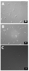
Figure A1
Figure A1
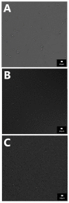
Photos of spermatozoa obtained with DIC using objectives with a high numeric aperture and various magnifications: (A) Leica DIC_100× NA 1.4 OIL 550 nm (total magnification 1000×); (B) Leica DIC_63× NA 1.4 OIL 550 nm (total magnification 630×); (C) Leica DIC_20× NA 1.0 OIL 550 nm zoom 3× (total magnification 600×). In either case, you can distinguish both large and small vacuoles in the sperm head.
Figure A2
Figure A2
Application of different filters with one objective, Leica DIC 20× NA 1.0 OIL (total magnification 100×): (A) 405 nm, (B) 550 nm, (C) 670 nm. The 405 and 550 nm show filters sufficient amount of detail. The 670 nm filter requires a more sensitive camera or more light.
Figure A3
Figure A3
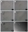
Photos of spermatozoa with HMC. Objectives used with various magnifications and numeric apertures: (A) 20× NA 0.3 (total magnification 200×)—sperm morphology details are not discernible, (B) 20× NA 0.45 (total magnification 200×)—larger vacuoles become visible, (C) 40× NA 0.60 (total magnification 400×)—larger vacuoles are visible at the sperm head, (D) 40× NA 0.95 (total magnification 400×)–vacuoles are visible even though the optimal Hoffman contrast was not achieved, (E) 60× NA 0.70 (total magnification 900×)—larger vacuoles are visible at the sperm head, (F) 60× NA 0.95 (total magnification 600×)–vacuoles are visible along with the correct spatial illusion.
Figure A4
Figure A4
Photos of spermatozoa with different modulation contrasts with an objective NA between 0.45 and 0.6 and a condenser NA between 0.4 and 0.6, no filter. (A) Hoffman modulation contrast—objective Nikon ELWD 20× NA 0.45, condenser NA 0.6, (total magnification 200×), (B) Hoffman modulation contrast—objective Nikon ELWD 40× NA 0.55, condenser NA 0.6, (total magnification 1000×) optical magnification 2.5 times within ocular tube head (T-TD), (C) Hoffman modulation contrast—objective Nikon ELWD 40× NA 0.55, condenser NA 0.6, (total magnification 400×), (D) Hoffman modulation contrast—objective Nikon ELWD 20× NA 0.45, condenser NA 0.6, (total magnification 500×), (E) relief contrast—objective Olympus LUCPlanFLN 20× NA 0.45, condenser NA 0.5, (total magnification 200×), (F) relief contrast—objective Olympus LUCPlanFLN 40× NA 0.60, condenser NA 0.5, (total magnification 400×), (G) PlasDIC—Zeiss, objective LD Plan-Neofluar 20× NA 0.4 condenser NA 0.55, (total magnification 200×), (H) PlasDIC—Zeiss, objective LD Plan-Neofluar 40× NA 0.6 condenser NA 0.55, (total magnification 400×).
Figure 1
Figure 1
Inverse relationship of numerical aperture and working distance.

References

    1. Palermo G., Joris H., Devroey P., van Steirteghem A.C. Pregnancies after intracytoplasmic injection of single spermatozoon into an oocyte. Lancet. 1992;340:17–18. doi: 10.1016/0140-6736(92)92425-F. - DOI - PubMed
    1. Ferraretti A.P., Nygren K., Andersen A.N., de Mouzon J., Kupka M., Calhaz-Jorge C., Wyns C., Gianaroli L., Goossens V., European IVF-Monitoring Consortium (EIM), for the European Society of Human Reproduction and Embryology (ESHRE) Trends over 15 years in ART in Europe: An analysis of 6 million cycles. Hum. Reprod. Open. 2017;2017:hox012. doi: 10.1093/hropen/hox012. - DOI - PMC - PubMed
    1. Alukal J.P., Lamb D.J. Intracytoplasmic sperm injection (ICSI)—What are the risks? Urol. Clin. N. Am. 2008;35:277–288. doi: 10.1016/j.ucl.2008.01.004. - DOI - PMC - PubMed
    1. Wong M.Y.W., Ledger W.L. Is ICSI Risky? Obstet. Gynecol. Int. Hindawi. 2013;2013:473289. doi: 10.1155/2013/473289. - DOI - PMC - PubMed
    1. Bartoov B., Berkovitz A., Eltes F. Selection of spermatozoa with normal nuclei to improve the pregnancy rate with intracytoplasmic sperm injection. N. Engl. J. Med. 2001;345:1067–1068. doi: 10.1056/NEJM200110043451416. - DOI - PubMed

Publication types

LinkOut - more resources