. 2022 Jan 3;12(1):63.
doi: 10.3390/life12010063.

Loss of Corticostriatal Mu-Opioid Receptors in α-Synuclein Transgenic Mouse Brains

Affiliations

Loss of Corticostriatal Mu-Opioid Receptors in α-Synuclein Transgenic Mouse Brains

Jessica Grigoletto et al. Life (Basel). .

Abstract

Ultrastructural, neurochemical, and molecular alterations within the striatum are associated with the onset and progression of Parkinson's disease (PD). In PD, the dopamine-containing neurons in the substantia nigra pars compacta (SNc) degenerate and reduce dopamine-containing innervations to the striatum. The loss of striatal dopamine is associated with enhanced corticostriatal glutamatergic plasticity at the early stages of PD. However, with disease progression, the glutamatergic corticostriatal white matter tracts (WMTs) also degenerate. We analyzed the levels of Mu opioid receptors (MORs) in the corticostriatal WMTs, as a function of α-Synuclein (α-Syn) toxicity in transgenic mouse brains. Our data show an age-dependent loss of MOR expression levels in the striatum and specifically, within the caudal striatal WMTs in α-Syn tg mouse brains. The loss of MOR expression is associated with degeneration of the myelinated axons that are localized within the corticostriatal WMTs. In brains affected with late stages of PD, we detect evidence confirming the degeneration of myelinated axons within the corticostriatal WMTs. We conclude that loss of corticostriatal MOR expression is associated with degeneration of corticostriatal WMT in α-Syn tg mice, modeling PD.

Keywords: corticostriatal glutamatergic axons; mu-opioid receptors (MOR); white matter tracts (WMTs); α-Synuclein.


Conflict of interest statement

The authors declare no conflict of interest.

Figures

Figure 1
Figure 1
Lower MOR levels in corticostriatal WMT of A53T α-Syn tg mouse brains. (A). A lower magnification of a coronal mouse brain section, stained with Luxol Fast blue, showing the striatum. CC, corpus callosum; LV, lateral ventricle; CPu, caudate putamen. WMTs are shown as dark spots over the lightly stained striatal grey matter in the CPu. Bar = 0.5 mm. (B). A semi-thick A53T α-Syn mouse brain section (1 μm), stained with methylene blue, showing cross-sectioned axons in a WMT. Bar = 10 μm. (C). Paraffin-embedded coronal sections (6 μm) of α-Syn-/- (C57BL/6JOlaHsd) and A53T α-Syn mouse brains at 2 months of age, containing the dorsal striatum (representative images). Sections immunoreacted either with anti-MOR, anti-NF-200, anti-MBP, or anti calbindin 28 (CB-28) antibodies. The consecutive section of the α-Syn-/- brain were stained with the corresponding secondary antibody as a control (no 1°). Bar = 50 μm. (D). The relative WMTs immunoreactive signal of MOR or MBP normalized to the signal obtained for NF-200. Total CB-28 signal in striatal matrix normalized to NF200 signal. Mean ± SD of A53T mice at 2, 8, and 12 months of age. The horizontal bar represents the corresponding age-matched control mice, set at 100%. n = 4–5 brains for each genotype and each age group; 3–6 fields per brain; t-test with Bonferroni correction, * p < 0.017.
Figure 2
Figure 2
MOR levels are lower in the striatum of Thy-1 WT α-Syn tg mouse brains. (A). Samples of membrane fractions P10 or P25, or supernatant S25 (30 μg protein) obtained from tissue punches containing the striatum of Thy-1 α-Syn and control mouse brains at 2 and 10 months of age analyzed by Western blotting and immunoreacted with anti-MOR or anti-Na+/K+ ATPase antibodies. The original western blot images are provided in the Supplementary material Figure S1 (mouse brain samples at 2 months of age) and Figure S2 (mouse brain samples at 10 months of age). (B). Graph showing quantitation of blots obtained in (A) mean ± SD of n = 4 mice. *, <0.05, t-test. (C). Western blot as in (A), the soluble S25 fraction immunoreacted with anti-CB-28 or anti β-actin antibodies. The original western blot images are provided in the Supplementary material Figure S3 (left side shows mouse brain samples at 2 months of age; the right side shows mouse brain samples at 10 months of age).
Figure 3
Figure 3
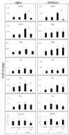
Lower mRNA levels of MOR in striatum and substantia nigra of A53T α-Syn tg mouse brains. Graphs showing qPCR values, detected following RNA extraction from tissue punches containing the striatum or SNc of A53T and age-matched control mouse brains, at 2 or 12–14 months of age (n = 4–5 mouse brains in each genotype and age group, tested in triplicates). The detected mRNA levels are normalized to the levels of 18S in the same sample. *, p < 0.007, t-test with Bonferroni correction for multiple comparisons.
Figure 4
Figure 4
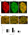
Axonal degeneration within caudal glutamatergic WMTs in progressive PD. (A). IHC of caudal WMTs in a human brain affected with early PD (male, 72 years, unified stage IIb). Brain section co-immunoreacted with anti-MBP and anti-SMI-32 antibodies. (B). IHC of a control human (male, 84 years); an early PD brain (as in A), and an advanced PD brain (male 75 years, Braak stage 5). Brain sections co-immunoreacted with anti-MBP and anti-SMI-32 antibodies. A representative brain is shown out of n = 3 in each group. Bar = 20 μm. (C). Quantitation of the signal as in (B) mean ± SD of n = 3 brains, 8–10 WMTs.

References

    1. Huston J.P. In: Hand Book of Basal Ganglia Structure and Function. 2nd ed. Steiner H., Tseng K.Y., editors. Volume 24. Elsevier; Cambridge, MA, USA: 2016. pp. 3–32. - DOI
    1. Haber S.N. Corticostriatal circuitry. Dialogues Clin. Neurosci. 2016;18:7–21. - PMC - PubMed
    1. Arkadir D., Bergman H., Fahn S. Redundant dopaminergic activity may enable compensatory axonal sprouting in Parkinson disease. Neurology. 2014;82:1093–1098. doi: 10.1212/WNL.0000000000000243. - DOI - PubMed
    1. Villalba R.M., Smith Y. Loss and remodeling of striatal dendritic spines in Parkinson’s disease: From homeostasis to maladaptive plasticity? J. Neural Transm. 2018;125:431–447. doi: 10.1007/s00702-017-1735-6. - DOI - PMC - PubMed
    1. Sgroi S., Tonini R. Opioidergic Modulation of Striatal Circuits, Implications in Parkinson’s Disease and Levodopa Induced Dyskinesia. Front Neurol. 2018;9:524. doi: 10.3389/fneur.2018.00524. - DOI - PMC - PubMed