. 2022 Jan 10;12(1):93.
doi: 10.3390/life12010093.

Overexpression of BQ323636.1 Modulated AR/IL-8/CXCR1 Axis to Confer Tamoxifen Resistance in ER-Positive Breast Cancer

Affiliations

Overexpression of BQ323636.1 Modulated AR/IL-8/CXCR1 Axis to Confer Tamoxifen Resistance in ER-Positive Breast Cancer

Ho Tsoi et al. Life (Basel). .

Erratum in

Abstract

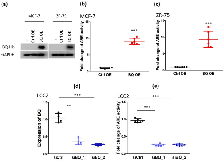
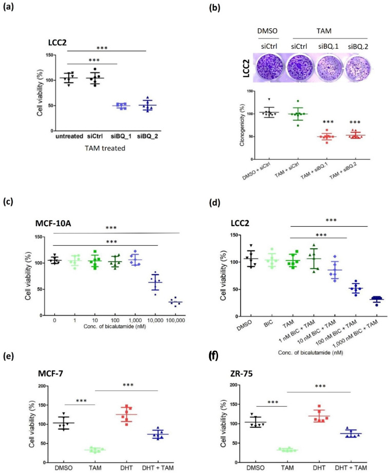
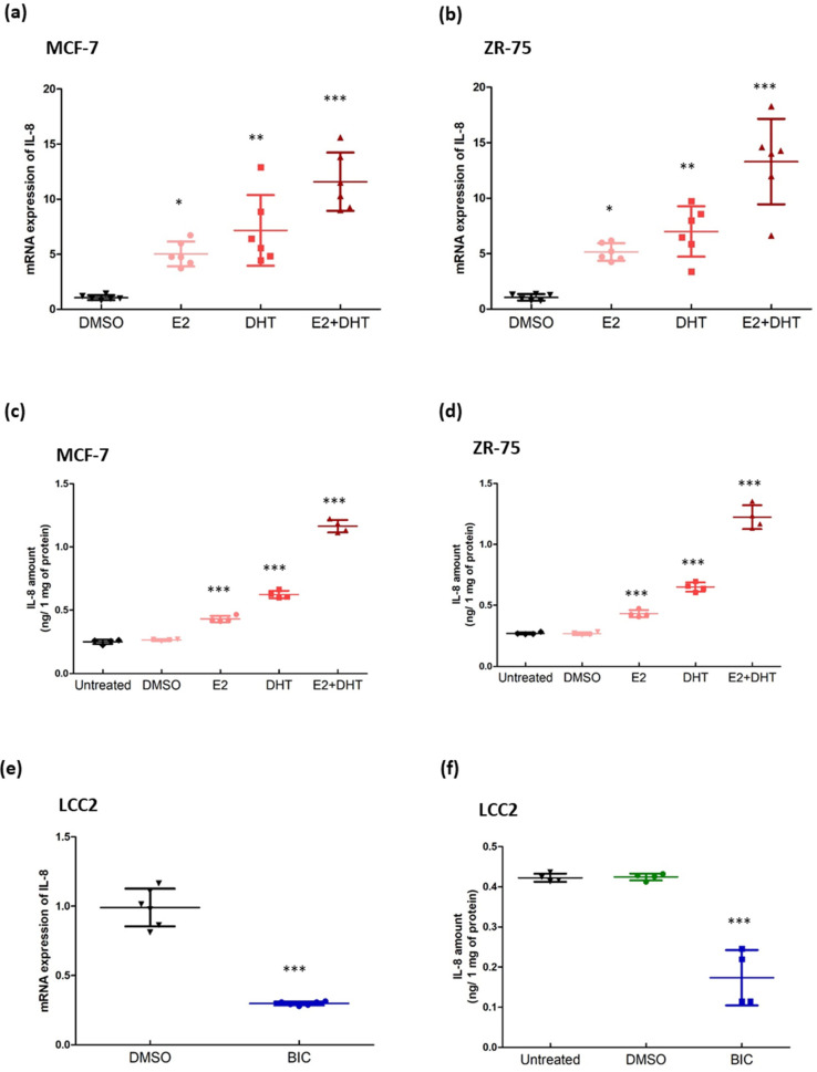
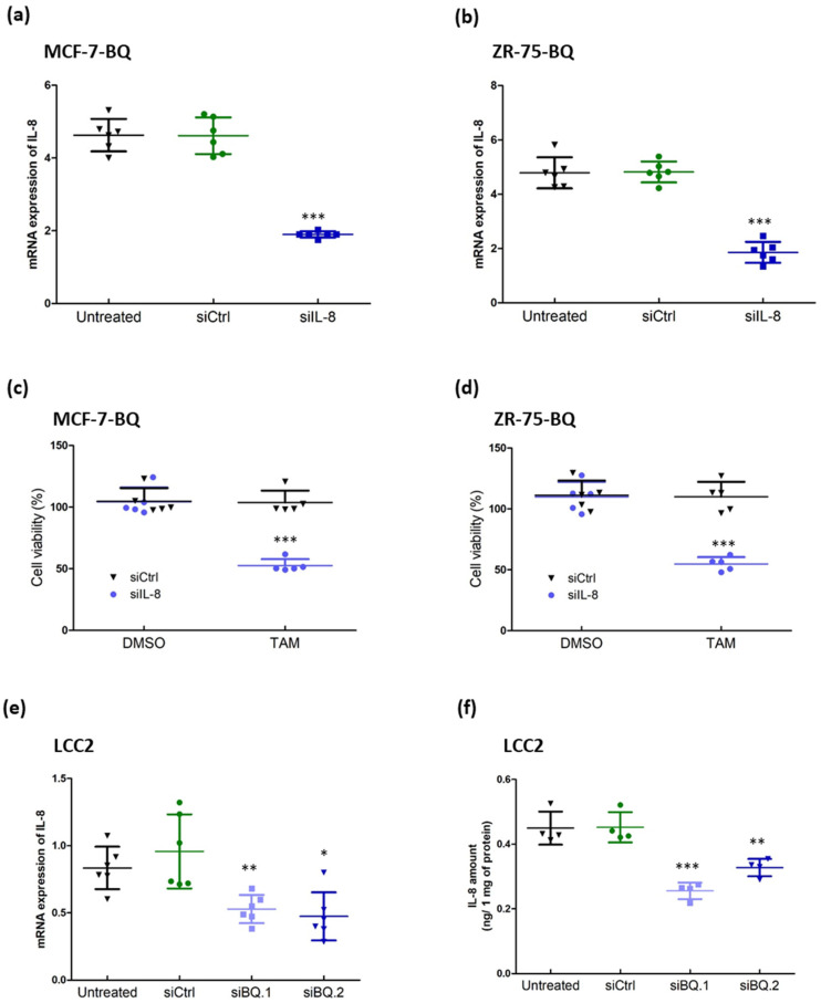
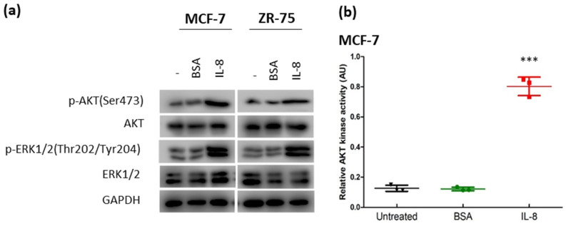
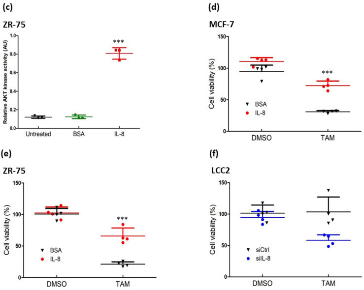
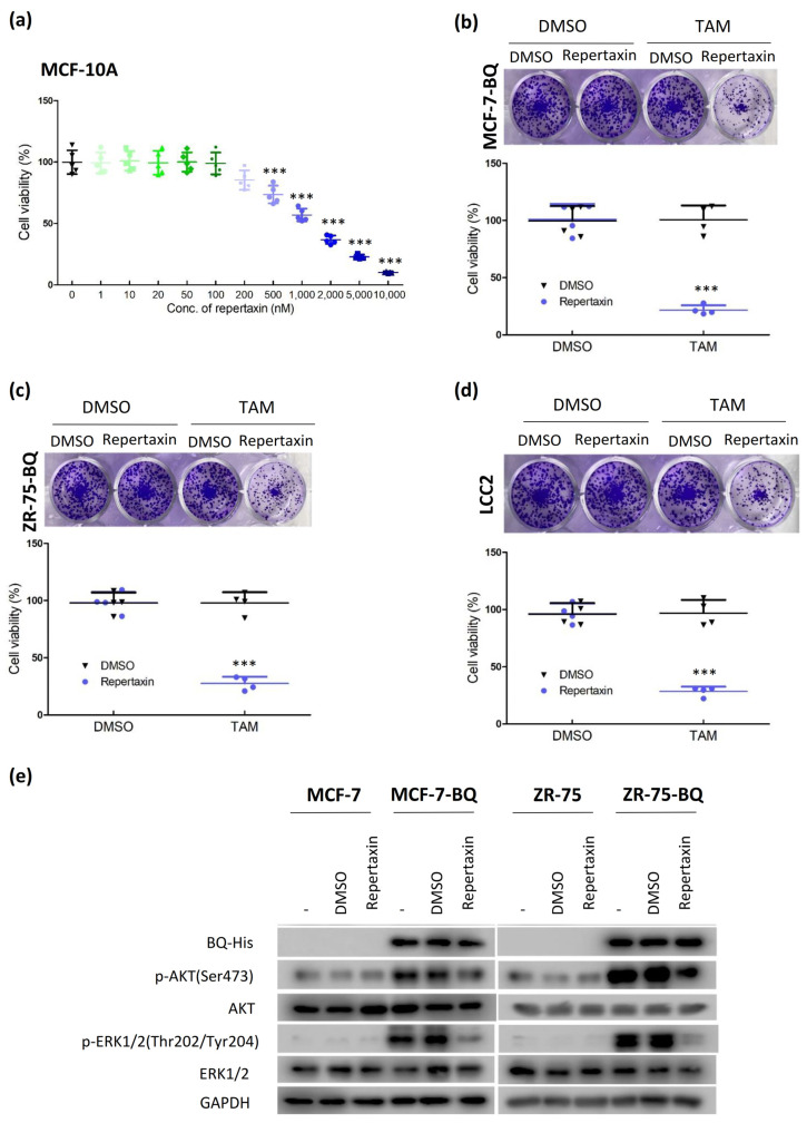
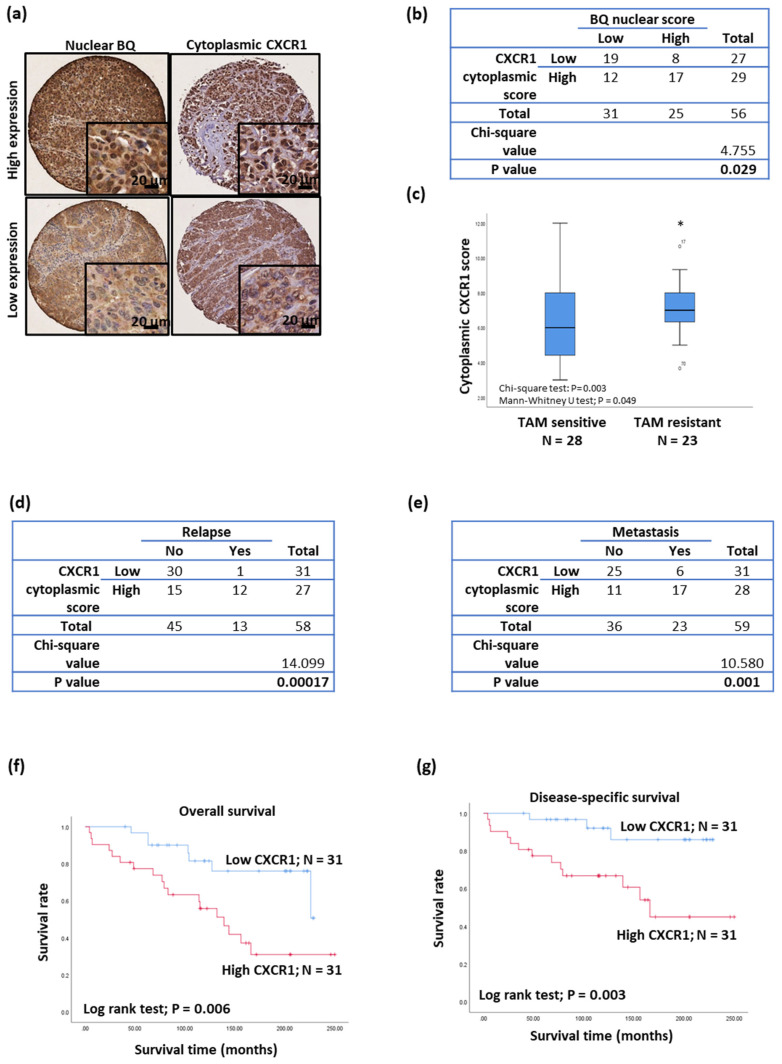
NCOR2 is a co-repressor for estrogen receptor (ER) and androgen receptor (AR). Our group previously identified a novel splice variant of NCOR2, BQ323636.1 (BQ), that mediates tamoxifen resistance via interference of NCOR2 repression on ER. Luciferase reporter assay showed BQ overexpression could enhance the transcriptional activity of androgen response element (ARE). We proposed that BQ employs both AR and ER to confer tamoxifen resistance. Through in silico analysis, we identified interleukin-8 (IL-8) as the sole ERE and ARE containing gene responsiveness to ER and AR activation. We confirmed that BQ overexpression enhanced the expression of IL-8 in ER+ve breast cancer cells, and AR inhibition reduced IL-8 expression in the BQ overexpressing cell lines, suggesting that AR was involved in the modulation of IL-8 expression by BQ. Moreover, we demonstrated that IL-8 could activate both AKT and ERK1/2 via CXCR1 to confer tamoxifen resistance. Targeting CXCR1/2 by a small inhibitor repertaxin reversed tamoxifen resistance of BQ overexpressing breast cancer cells in vitro and in vivo. In conclusion, BQ overexpression in ER+ve breast cancer can enhance IL-8 mediated signaling to modulate tamoxifen resistance. Targeting IL-8 signaling is a promising approach to overcome tamoxifen resistance in ER+ve breast cancer.

Keywords: BQ323636.1; CXCR1; androgen receptor; breast cancer; interleukin-8; tamoxifen resistance.


Conflict of interest statement

The authors declare no conflict of interest.

Figures

Figure 1
Figure 1
Overexpression of BQ323636.1 (BQ) can modulate the activity of androgen receptor (AR) in breast cancer cells. (a) Western blot was employed to confirm BQ ectopic expression in stable BQ overexpressing MCF-7 and ZR-75 cell lines. GAPDH was used as the loading control. Overexpression of BQ could enhance the transcriptional activity of AR in (b) MCF-7 and (c) ZR-75. Luciferase reporter assay with androgen response element (ARE) was employed. Results were shown as mean ± SD from 6 independent experiments. Student’s t-test was employed to determine statistical significance. *** represents p < 0.001. (d) Knockdown efficiency of siRNAs against BQ. LCC2 was transfected with 25 µM of non-targeting siRNA (siCtrl), siBQ.1 or siBQ.2. qPCR was performed 72 h post-transfection. Actin was used as the internal control. Results were shown as mean ± SD from 4 independent experiments. One-way ANOVA was employed. Bonferroni’s multiple comparison test was employed to determine the significance between 2 groups. ** and *** represent p < 0.01 and p < 0.001 respectively. (e) Knockdown of BQ could reduce AR activity in LCC2. Luciferase reporter assay with ARE was used. Results were shown as mean ± SD from 6 independent experiments. One-way ANOVA was employed. Bonferroni’s multiple comparison test was employed to determine the significance between 2 groups. *** represents p < 0.001.
Figure 2
Figure 2
Inhibition of AR could reverse tamoxifen resistance. (a) Knockdown of BQ could recover tamoxifen sensitivity as revealed by MTT assay. LCC2 was transfected with 25 µM of non-targeting siRNA (siCtrl), siBQ.1 or siBQ.2. 48 h post-transfection, the cells were treated with 4 µM of tamoxifen (4-OHT; TAM) for 96 h. MTT assay was employed to determine cell viability. Results were shown as mean ± SD from 6 independent experiments. (b) Knockdown of BQ could recover tamoxifen sensitivity as revealed by clonogenic assay. 48 h post-transfection, the cells were treated with 4 µM of tamoxifen (4-OHT; TAM) for 14 days. 0.01% crystal violet was used to stain the cells. Results were shown as mean ± SD from 9 independent experiments. (c) The effect of AR antagonist bicalutamide on cell viability of MCF-10A. The cells were treated with different concentrations of bicalutamide for 96 h. MTT assay was employed to determine cell viability. Results were shown as mean ± SD from 6 independent experiments. (d) Dosage dependent effect of bicalutamide on reversing tamoxifen resistance in LCC2. The cells were treated with4 µM of tamoxifen (4-OHT; TAM) and different concentrations of bicalutamide (BIC) for 96 h. MTT assay was employed to determine cell viability. Results were shown as mean ± SD from 6 independent experiments. Activation of AR could decrease the efficacy of tamoxifen in (e) MCF-7 and (f) ZR-75. The cells were treated with 4 µM of tamoxifen (4-OHT; TAM) 0.1 nM of dihydrotestosterone (DHT; androgen) for 96 h. MTT assay was employed to determine cell viability. Results were shown as mean ± SD from 6 independent experiments. One-way ANOVA was employed. Bonferroni’s multiple comparison test was employed to determine the significance between 2 groups. *** represent p < 0.001.
Figure 3
Figure 3
Modulating the activity of AR could interfere with the expression of IL-8. The effect of estrogen and androgen on the expression of IL-8 in (a) MCF-7 and (b) ZR-75. The cells were treated with 1 nM of Estradiol (E2; estrogen) and/or 0.1 nM of dihydrotestosterone (DHT; androgen) for 24 h. qPCR was performed. Actin was used as the internal control. Results were shown as mean ± SD from 6 independent experiments. One-way ANOVA was employed. Bonferroni’s multiple comparison test was employed to determine the significance between DMSO and any of the treatment groups. (c,d) ELISA was performed to confirm the effect of 1 nM of E2 and 0.1 nM of DHT on the production of IL-8. Culture medium was collected after 24 h of the treatment. ELISA was performed to determine the amount of IL-8 in the medium. Results were shown as mean ± SD from 4 independent experiments. One-way ANOVA was employed. Bonferroni’s multiple comparison test was employed to determine the significance between untreated and any of the treatment groups. Suppression of AR could reduce the (e) mRNA expression and (f) protein production of IL-8 in LCC2. The cells were treated with 1 µM of bicalutamide (BIC; AR antagonist) for 48 h. qPCR was performed to determine mRNA. Results were shown as mean ± SD from 4 independent experiments. Student’s t-test was employed to determine statistical significance. ELISA was performed to evaluate the production of IL-8 in the culture medium. Results were shown as mean ± SD from 4 independent experiments. One-way ANOVA was employed. Bonferroni’s multiple comparison test was employed to determine the significance between untreated and BIC treated groups. *, **, and *** represent p < 0.05, p < 0.01 and p < 0.001.
Figure 4
Figure 4
Modulating the expression of IL-8 could alter the response to tamoxifen. Knockdown efficiency of siRNA against IL-8 in (a) MCF-7-BQ and (b) ZR-75-BQ. The cells were treated with 25 µM of non-targeting siRNA (siCtrl) or IL-8 specific siRNA (siIL-8). qPCR was performed 48 h post-transfection. Actin was used as the internal control. Results were shown as mean ± SD from 6 independent experiments. One-way ANOVA was employed. Bonferroni’s multiple comparison test was employed to determine the significance between untreated and siIL-8 treated groups. Knockdown of IL-8 could reverse tamoxifen resistance in (c) MCF-7-BQ and (d) ZR-75-BQ. The cells were transfected with siCtrl and siIL-8. 4 µM of 4-OHT (TAM) was used after 48 h of the transfection. MTT assay was performed to determine cell viability after 72 h of TAM treatment. Results were shown as mean ± SD from 5 independent experiments. Student’s t-test was employed to determine statistical significance between siCtrl and siIL-8 treated groups. Knockdown of BQ could reduce the (e) mRNA expression and (f) protein expression of IL-8 in LCC2. LCC2 cells were treated with the siRNAs. qPCR was performed to determine the mRNA level of IL-8, 48 h post-transfection. Results were shown as mean ± SD from 6 independent experiments. ELISA was performed to determine the amount of IL-8 in the culture medium. Results were shown as mean ± SD from 4 independent experiments. One-way ANOVA was employed. Bonferroni’s multiple comparison test was employed to determine the significance between untreated and siRNAs treated groups. *, **, and *** represent p < 0.05, p < 0.01 and p < 0.001.
Figure 5
Figure 5
Treatment of IL-8 could induce tamoxifen resistance. (a) IL-8 could activate AKT and ERK1/2 in MCF-7 and ZR-75 cells. The cells were treated with 5 ng/mL of IL-8. Proteins were harvested 24 h post-treatment. Western blot was employed to determine the expression of the candidate proteins. GAPDH was the loading control. The treatment of IL-8 could enhance AKT kinase activity in (b) MCF-7 and (c) ZR-75. The cells were treated with 5 ng/mL of IL-8. AKT kinase activity assay was performed after 24 h of the treatment. Results were shown as mean ± SD from 3 independent experiments. One-way ANOVA was employed. Bonferroni’s multiple comparison test was employed to determine the significance between untreated and IL-8 treated groups. IL-8 treatment could enhance the tolerance to tamoxifen in (d) MCF-7 and (e) ZR-75. The cells were co-treated with 4 µM of 4-OHT (TAM) and 5 ng/mL of IL-8 or BSA for 96 h. MTT assay was employed to determine cell viability. Results were shown as mean ± SD from 4 independent experiments. Student’s t-test was employed to determine statistical significance between BSA, and IL-8 treated groups. (f) Knockdown of IL-8 could reduce tamoxifen resistance in LCC2. The cells were transfected with 25 µM of siCtrl or siIL-8. The cells were treated with 4 µM of TAM after 48 h of the transfection. MTT assay was performed after 96 h of TAM treatment. Results were shown as mean ± SD from 4 independent experiments. Student’s t-test was employed to determine statistical significance between siCtrl and siIL-8 treated groups. *** represents p < 0.001.
Figure 5
Figure 5
Treatment of IL-8 could induce tamoxifen resistance. (a) IL-8 could activate AKT and ERK1/2 in MCF-7 and ZR-75 cells. The cells were treated with 5 ng/mL of IL-8. Proteins were harvested 24 h post-treatment. Western blot was employed to determine the expression of the candidate proteins. GAPDH was the loading control. The treatment of IL-8 could enhance AKT kinase activity in (b) MCF-7 and (c) ZR-75. The cells were treated with 5 ng/mL of IL-8. AKT kinase activity assay was performed after 24 h of the treatment. Results were shown as mean ± SD from 3 independent experiments. One-way ANOVA was employed. Bonferroni’s multiple comparison test was employed to determine the significance between untreated and IL-8 treated groups. IL-8 treatment could enhance the tolerance to tamoxifen in (d) MCF-7 and (e) ZR-75. The cells were co-treated with 4 µM of 4-OHT (TAM) and 5 ng/mL of IL-8 or BSA for 96 h. MTT assay was employed to determine cell viability. Results were shown as mean ± SD from 4 independent experiments. Student’s t-test was employed to determine statistical significance between BSA, and IL-8 treated groups. (f) Knockdown of IL-8 could reduce tamoxifen resistance in LCC2. The cells were transfected with 25 µM of siCtrl or siIL-8. The cells were treated with 4 µM of TAM after 48 h of the transfection. MTT assay was performed after 96 h of TAM treatment. Results were shown as mean ± SD from 4 independent experiments. Student’s t-test was employed to determine statistical significance between siCtrl and siIL-8 treated groups. *** represents p < 0.001.
Figure 6
Figure 6
Treatment of CXCR1/2 inhibitor, repertaxin could reduce tamoxifen resistance. (a) The dosage-dependent effect of repertaxin on cell viability of MCF-10A. Non-cancerous breast epithelial cell line MCF-10A was used. The cells were treated with different concentrations of repertaxin for 96 h. MTT assay was performed. Results were shown as mean ± SD from 5 independent experiments. One-way ANOVA was employed. Bonferroni’s multiple comparison test was employed to determine the significance between 0 nM and other concentrations. Repertaxin could reduce tamoxifen resistance in (b) MCF-7-BQ, (c) ZR-75-BQ and (d) LCC2. The cells were co-treated with 4 µM of 4-OHT and 0.1 µM of repertaxin for 14 days. Clonogenic assay was performed. 0.01% crystal violet was used to stain the colonies. Results were shown as mean ± SD from 4 independent experiments. Student’s t-test was employed to determine statistical significance between DMSO and repertaxin treated groups. *** represents p < 0.001. (e) Repertaxin could suppress AKT and ERK1/2 activation on BQ overexpressing cells. MCF-7, MCF-7-BQ, ZR-75 and ZR-75-BQ cells were treated with 0.1 µM of repertaxin for 48 h. Western blot was used to determine the expression of the protein candidates. GAPDH was used as the loading control.
Figure 7
Figure 7
Repertaxin could reverse tamoxifen resistance in vivo. (a) ZR-75-BQ cell line was employed to establish xenografts. The cells were implanted onto the mammary fat pad of nude mice. The mice received saline, tamoxifen (4-OHT; 500 mg; twice per week), repertaxin (15 mg/Kg; twice per week), tamoxifen + repertaxin (500 mg of 4-OHT + 7.5 mg/Kg of repertaxin; twice per week) and tamoxifen + repertaxin (500 mg of 4-OHT + 15 mg/Kg of repertaxin; twice per week). Repertaxin was delivered by subcutaneous injection. After 4 weeks of treatment, tumors were harvested. (b) The photo showed the effect of different treatments on tumor size. Results were shown as mean ± SD from 4 independent tumors. Two-way ANOVA was performed. Bonferroni’s multiple comparison test was employed to determine the significance between saline and other treatment groups at each time point. ** and *** represent p < 0.01 and p < 0.001, respectively. (c) Treatment of repertaxin could reduce the levels of activated AKT and ERK1/2 in the tumors. Proteins were harvested from the tumors. Western blot was performed to analyze the expression of the indicated protein candidates in 3 of the independent tumors. GAPDH was used as the loading control.
Figure 8
Figure 8
Clinical significance of CXCR1 in breast cancer. (a) Immunohistochemistry of BQ and CXCR2 was performed on primary ER+ve breast tumor on TMA. (b) Chi-square test to determine the correlation between nuclear BQ and cytoplasmic CXCR1. (c) Tamoxifen resistance was associated with high expression of cytoplasmic CXCR1. Chi-square test and Mann–Whitney U test were employed. * represents p < 0.05. Chi-square test to determine the correlation of cytoplasmic CXCR1 with (d) relapse and (e) metastasis. High expression of CXCR1 was associated with poorer (f) overall survival and (g) disease-specific survival in ER+ve breast cancer. Log-rank test was employed.

References

    1. Sung H., Ferlay J., Siegel R.L., Laversanne M., Soerjomataram I., Jemal A., Bray F. Global Cancer Statistics 2020: GLOBOCAN Estimates of Incidence and Mortality Worldwide for 36 Cancers in 185 Countries. CA Cancer J. Clin. 2021;71:209–249. doi: 10.3322/caac.21660. - DOI - PubMed
    1. Sørlie T., Perou C.M., Tibshirani R., Aas T., Geisler S., Johnsen H., Børresen-Dale A.L. Gene expression patterns of breast carcinomas distinguish tumor subclasses with clinical implications. Proc. Natl. Acad. Sci. USA. 2001;98:10869–10874. doi: 10.1073/pnas.191367098. - DOI - PMC - PubMed
    1. Rozeboom B., Dey N., De P. ER+ metastatic breast cancer: Past, present, and a prescription for an apoptosis-targeted future. Am. J. Cancer Res. 2019;9:2821–2831. - PMC - PubMed
    1. Kulkoyluoglu E., Madak-Erdogan Z. Nuclear and extranuclear-initiated estrogen receptor signaling crosstalk and endocrine resistance in breast cancer. Steroids. 2016;114:41–47. doi: 10.1016/j.steroids.2016.06.007. - DOI - PubMed
    1. Katzenellenbogen J.A., Mayne C.G., Katzenellenbogen B.S., Greene G.L., Chandarlapaty S. Structural underpinnings of oestrogen receptor mutations in endocrine therapy resistance. Nat. Rev. Cancer. 2018;18:377–388. doi: 10.1038/s41568-018-0053-0. - DOI - PMC - PubMed