Review
. 2022 Jan 10;23(2):727.
doi: 10.3390/ijms23020727.

Role of Palatine Tonsil and Epipharyngeal Lymphoid Tissue in the Development of Glomerular Active Lesions (Glomerular vasculitis) in Immunoglobulin A Nephropathy

Affiliations
Review

Role of Palatine Tonsil and Epipharyngeal Lymphoid Tissue in the Development of Glomerular Active Lesions (Glomerular vasculitis) in Immunoglobulin A Nephropathy

Osamu Hotta et al. Int J Mol Sci. .

Abstract

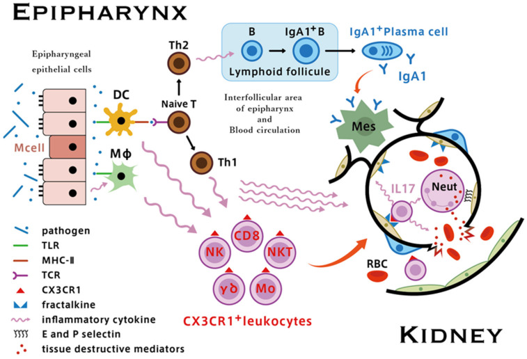
Hematuria is an essential symptom of immunoglobulin A nephropathy (IgAN). Although the etiology of hematuria in IgAN has not been fully elucidated, it is thought that the rupture of the glomerular basement membranes caused by intra-capillary leukocyte influx, so-called glomerular vasculitis, is the pathological condition responsible for severe hematuria. Glomerular vasculitis are active lesions that exist in the glomeruli of acute phase IgAN and it is important because it is suspected to make the transition to segmental glomerular sclerosis (SGS) as a repair scar lesion in the chronic phase, and the progression of SGS would eventually lead to glomerular obsolescence. Worsening of hematuria concomitant with acute pharyngitis is common in patients with IgAN; therefore, elucidating the relationship between the immune system of Waldeyer's ring, including the palatine tonsil and epipharyngeal lymphoid tissue, and the glomerular vasculitis may lead to understanding the nature of IgAN. The epipharynx is an immunologically activated site even under normal conditions, and enhanced activation of innate immunity is likely to occur in response to airborne infection. Hyperactivation of innate immunity via upregulation of Toll-like receptors in the interfollicular area of the palatine tonsil and epipharyngeal lymphoid tissue, followed by enhanced fractalkine/CX3CR1 interactions, appears to play an important role in the development of glomerular vasculitis in IgAN. As latent but significant epipharyngitis is present in most patients with IgAN, it is plausible that acute upper respiratory infection may contribute as a trigger for the innate epipharyngeal immune system, which is already upregulated in a chronically inflamed environment. Given that epipharyngitis and its effects on IgAN are not fully understood, we propose that the so-called "epipharynx-kidney axis" may provide an important focus for future research.

Keywords: IgA nephropathy; chronic epipharyngitis; chronic tonsillitis; epipharynx–kidney axis; glomerular vasculitis.


Conflict of interest statement

The authors do not have any financial relationships that may lead to a conflict of interest in relation to the submitted manuscript.

Figures

Figure 1
Figure 1
Mesangial expansion in immunoglobulin A nephropathy (IgAN) with massive IgA deposition. In cases with massive IgA deposition, pronounced hemispherical deposits are often observed (left PAS). Regardless of the extent of mesangium proliferation in response to IgA deposition, it is located in the extra-capillary area (brown) and did not occlude the glomerular vascular cavity (right, PAM).
Figure 2
Figure 2
The glomerular sclerosing process following glomerular vasculitis. Following glomerular vasculitis (a), segmental glomerular sclerosis develops as a healing scar lesion (b). The accumulation and expansion of segmental glomerular sclerosis lesions (c) ultimately lead to glomerular obsolescence (d). PAS, original magnification ×400.
Figure 3
Figure 3
Progression factors in the time course of immunoglobulin A nephropathy (IgAN). Glomerular vasculitis is the main progression factor in the early stages of IgAN. Meanwhile, common pathways, such as glomerular hypertension, ischemia, and proteinuria-related tubulointerstitial injury, are the main contributing progression factors in the later stage of IgAN. Glomerular vasculitis can be extinguished by an effective therapeutic intervention, such as steroid pulse and tonsillectomy, but complete blocking of the common pathway is impossible for now. Therefore, there is a “point of no remission” in the time course to achieve remission or cure of IgAN.
Figure 4
Figure 4
Lymphoepithelial symbiosis in palatine tonsils. The tonsillar crypt epithelium and tonsil parenchyma are mixed, which is called “lymphoepithelial symbiosis.” Reticular epithelial cells (cytokeratin positive brown cells)-fold lymphocytes (blue). Immunoperoxidase staining with anti-cytokeratin antibody (brown), original magnification ×100.
Figure 5
Figure 5
Reducing lymphoepithelial symbiosis and the progression of immunoglobulin A nephropathy (IgAN). The area of non-reticulated crypts (non-lymphoepithelial symbiosis areas) expands with the progression of IgAN. Representative cases: (a) CKD stage 2, disease period of 2 years, and (b) CKD stage 5, disease period of 30 years. The area of non-reticulated epithelia (red box) is restricted in the early stage of IgAN (a), whereas non-reticulation and squamous epithelialization are remarkable in ESKD IgAN (b). Stained with anti-cytokeratin antibody (brown), original magnification ×40.
Figure 6
Figure 6
T cell nodules in palatine tonsil. T cell nodules (red arrows) consisting of CD3+CD20-HLA-DR+ cells present in the interfollicular area of the palatine tonsil (original magnification ×40).
Figure 7
Figure 7
Role of palatine tonsils in the pathogenesis of immunoglobulin A nephropathy (IgAN). The palatine tonsil has a unique structure, namely, lymphoepithelial symbiosis, which functions as an induction site in the mucosal immune system. IgAN tonsils have characteristics such as failure of immune tolerance against indigenous bacteria and a hyperimmune response against bacteria-originated DNA. The overproduction of aberrantly glycosylated and polymeric IgA1 through BAFF and APRIL mediated T cell-independent pathways in lymphoid follicles in tonsils may be a source of mesangial IgA deposition. The increased number of T cell nodules co-localized with CD208+ dendritic cells in IgAN tonsils may be related to the enhanced expression of CX3CR1 in CD8+ cells and other types of lymphocytes with cytotoxic activity and the simultaneous upregulation of fractalkine/CX3CR1 interaction in distant glomeruli occurs, resulting in glomerular vasculitis.
Figure 8
Figure 8
Surface of the epipharynx. There is an abundance of lymphocytes in the submucosal area (left, hematoxylin and eosin staining, original magnification ×100). Many lymphocytes are located within ciliated epithelial cells (right, indirect immunoperoxidase staining with anti-CD3 antibody, original magnification ×100).
Figure 9
Figure 9
Putative mechanisms of the epipharynx–kidney axis causing glomerular vasculitis. In acute epipharyngitis, following pathogen recognition through TLRs, dendritic cells in the mucosa immediately produce proinflammatory cytokines. Simultaneously, infected epithelial cells and activated macrophages produce chemokines that upregulate the expression of CX3CR1 in monocytes, NK cells, CD8+ T cells, and γδ T cells. Circulating proinflammatory cytokines also enhance the expression of fractalkine and other adhesion molecules such as E- and P-selectins in glomerular endothelial cells. Upregulation of the fractalkine/CX3CR1 interaction and IL17 secreted by infiltrated glomerular CX3CR1+ leukocytes, which promotes the influx of neutrophils, leads to the development of Glomerular vasculitis and subsequent rupture of the glomerular capillary wall, which is clinically associated with worsening of hematuria [55].
Figure 10
Figure 10
Endoscopic findings of chronic epipharyngitis. The vasculature is clearly visualized in the image-enhanced mode using band-limited light under normal conditions (a). In contrast, the vasculature is not visible due to submucosal edema and congestion, and the surface of the epipharynx becomes uneven and paved depending on the severity of chronic epipharyngitis (bd). In a representative case of IgA nephropathy (eg), the vasculature is obscure, and the surface exhibits a folded and cobblestone-like appearance (e,f). It is prone to bleeding by abrasion with a cotton swab (arrow) (g). Standard mode (e,g) and enhanced mode using band-limited light (ad,f).
Figure 11
Figure 11
Schematic illustration of epipharyngeal abrasive therapy. A cotton swab soaked with 0.5% ZnCl2 solution was inserted through both nostrils and rubbed approximately 30 times on each side. Next, the entire epipharyngeal wall was scrubbed using a pharyngeal swab soaked in 0.5% ZnCl2 solution.
Figure 12
Figure 12
Effects of epipharyngeal abrasive therapy on hematuria in patients with IgAN with TSP resistant or relapsed course.
Figure 13
Figure 13
Effects of epipharyngeal abrasive therapy on hematuria in potential IgAN.
Figure 14
Figure 14
Treatment options based on the Glomerular vasculitis related pathologic conditions of IgAN. As Glomerular vasculitis and segmental glomerular sclerosis (SGS) differently coexist in IgAN, the key point of IgAN treatment is to estimate the element of Glomerular vasculitis and the element of the SGS from the findings of renal biopsy and urinalysis in each patient. It is also important to consider the limitation of histological assessment of Glomerular vasculitis by a limited amount of renal biopsy tissue. See the text for further details. RAS, renin-angiotensin system; SGLT2, sodium-glucose cotransporter 2; LPG, low-protein diet; EAT, epipharyngeal abrasive therapy.

References

    1. Suzuki K., Honda K., Tanabe K., Toma H., Nihei H., Yamaguchi Y. Incidence of latent mesangial IgA deposition in renal allograft donors in Japan. Kidney Int. 2003;63:2286–2294. doi: 10.1046/j.1523-1755.63.6s.2.x. - DOI - PubMed
    1. Bellur S.S., Troyanov S., Cook H.T., Roberts I.S., Working Group of International IgA Nephropathy Network and Renal Pathology Society Immunostaining findings in IgA nephropathy: Correlation with histology and clinical outcome in the Oxford classification patient cohort. Nephrol. Dial. Transplant. 2011;26:2533–2536. doi: 10.1093/ndt/gfq812. - DOI - PubMed
    1. Nicholls K., Walker R.G., Dowling J.P., Kincaid-Smith P. “Malignant” IgA nephropathy. Am. J. Kidney Dis. 1985;5:42–46. doi: 10.1016/S0272-6386(85)80134-7. - DOI - PubMed
    1. Kincaid-Smith P., Nicholls K., Birchall I. Polymorphs infiltrate glomeruli in mesangial IgA glomerulonephritis. Kidney Int. 1989;36:1108–1111. doi: 10.1038/ki.1989.308. - DOI - PubMed
    1. Tewari R., Nada R., Kaur M., Dudeja P., Rayat C.S., Sakhuja V., Joshi K. Correlates of hematuria on glomerular histology and electron microscopy in IgA nephropathy. Med. J. Armed Forces India. 2016;72:120–124. doi: 10.1016/j.mjafi.2016.01.005. - DOI - PMC - PubMed

MeSH terms