. 2022 Jan 14;23(2):866.
doi: 10.3390/ijms23020866.

Therapeutic CFTR Correction Normalizes Systemic and Lung-Specific S1P Level Alterations Associated with Heart Failure

Affiliations

Therapeutic CFTR Correction Normalizes Systemic and Lung-Specific S1P Level Alterations Associated with Heart Failure

Franziska E Uhl et al. Int J Mol Sci. .

Abstract

Heart failure (HF) is among the main causes of death worldwide. Alterations of sphingosine-1-phosphate (S1P) signaling have been linked to HF as well as to target organ damage that is often associated with HF. S1P's availability is controlled by the cystic fibrosis transmembrane regulator (CFTR), which acts as a critical bottleneck for intracellular S1P degradation. HF induces CFTR downregulation in cells, tissues and organs, including the lung. Whether CFTR alterations during HF also affect systemic and tissue-specific S1P concentrations has not been investigated. Here, we set out to study the relationship between S1P and CFTR expression in the HF lung. Mice with HF, induced by myocardial infarction, were treated with the CFTR corrector compound C18 starting ten weeks post-myocardial infarction for two consecutive weeks. CFTR expression, S1P concentrations, and immune cell frequencies were determined in vehicle- and C18-treated HF mice and sham controls using Western blotting, flow cytometry, mass spectrometry, and qPCR. HF led to decreased pulmonary CFTR expression, which was accompanied by elevated S1P concentrations and a pro-inflammatory state in the lungs. Systemically, HF associated with higher S1P plasma levels compared to sham-operated controls and presented with higher S1P receptor 1-positive immune cells in the spleen. CFTR correction with C18 attenuated the HF-associated alterations in pulmonary CFTR expression and, hence, led to lower pulmonary S1P levels, which was accompanied by reduced lung inflammation. Collectively, these data suggest an important role for the CFTR-S1P axis in HF-mediated systemic and pulmonary inflammation.

Keywords: cystic fibrosis transmembrane regulator; heart failure; inflammation; lung; sphingosine-1-phosphate.


Conflict of interest statement

The authors declare no conflict of interest.

Figures

Figure A1
Figure A1
Gene expression of sphingosine-1-phosphate generating enzymes (a) SphK1 and (b) SphK2, and S1P degrading enzymes (c) Sgpl, (d) Sgpp1 and (e) Sgpp2 in the lungs of sham and HF mice. N = 8. HFheart failure; S1Psphingosine-1-phosphate; SphKsphingosine kinase, SgplS1P lyase, SgppS1P phosphatase.
Figure 1
Figure 1
Impaired pulmonary cystic fibrosis transmembrane regulator expression during heart failure links to increased sphingosine-1-phosphate concentrations in the lung. (a) CFTR protein expression in lung tissue of HF mice and sham-operated controls determined with Western blotting. Inset showing representative CFTR protein expression pattern. (b) S1P levels in lung tissue of HF mice and sham-operated controls assessed by mass spectrometry. (c) Pulmonary S1P levels of CFTR mutant mice (dF508 mutation) and littermate controls assessed by mass spectrometry. Data are expressed as mean ± SEM, t-test where * denotes p ≤ 0.05. CFTRcystic fibrosis transmembrane regulator; dF508delta F508 CFTR mutant; HFheart failure; Ssham; S1Psphingosine-1-phosphate; WTwild-type.
Figure 2
Figure 2
Experimental timeline. Male C57Bl/6N WT mice were subjected to myocardial infarction by permanent left anterior descending coronary artery ligation. Ten weeks post-myocardial infarction, mice received daily intraperitoneal injections of C18 (3 mg/kg BW) or an equivalent volume of vehicle for two consecutive weeks. Tissue was harvested and subjected to different experimental procedures. BWbody weight; CFTRcystic fibrosis transmembrane regulator; WTwild-type.
Figure 3
Figure 3
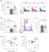
Correcting cystic fibrosis transmembrane regulator expression attenuates heart failure-associated pulmonary sphingosine-1-phosphate elevation. (a) Proportion of CFTR+ cells in the lungs of sham and HF mice after two weeks of vehicle or C18 treatment determined by flow cytometry. Representative zebra plots showing proportion of CFTR+ cells in lung tissue. (b) MFI quantification of CFTR+ cells in lung tissue of sham and HF mice after two weeks of vehicle or C18 treatment determined by flow cytometry and representative histograms. (c) Pulmonary S1P tissue concentrations of vehicle and C18-treated HF mice. The pink dotted line indicates sham levels. (d) Linear regression showing associations between the proportion of CFTR+ cells in lung tissue and pulmonary S1P levels. (e) Linear regression showing associations between the MFI of CFTR+ cells in lung tissue and pulmonary S1P levels. Data are expressed as mean ± SEM. In (a,b), one-way ANOVA with Tukey’s post hoc testing where * denotes p ≤ 0.05 between sham and HF + vehicle and & denotes p ≤ 0.05 between HF + vehicle and HF + C18; in (c) t-test where * denotes p ≤ 0.05; in (d,e), linear regression and Pearson’s correlation with exact r- and p-value computation. CFTRcystic fibrosis transmembrane regulator; HFheart failure; MFImedian fluorescence intensity; S1Psphingosine-1-phosphate.
Figure 4
Figure 4
Correction of cystic fibrosis transmembrane regulator expression normalizes sphingosine-1-phosphate plasma levels with implications for systemic inflammation. (a) Plasma S1P concentrations during HF disease progression. (b) Comparison of S1P plasma levels between vehicle- and C18-treated HF mice. The pink dotted line indicates sham levels. (c) Frequency of splenic S1P1+ CD3+ T-cells assessed by flow cytometry in vehicle- and C18-treated HF mice. The pink dotted line indicates sham levels. Representative histograms showing the proportion of S1P1+ CD3+ T-cells in spleen tissue of HF mice after two weeks of vehicle (pink) or C18 (blue) treatment compared to their respective FMO controls (gray). In (a) two-way ANOVA where * denotes p ≤ 0.05 between baseline and different timepoints after Sidak post hoc testing; & denotes p ≤ 0.05 between sham and HF after Tukey’s post hoc testing; In (b,c), t-test where & denotes p ≤ 0.05. CDcluster of differentiation; FMOfluorescence minus one, HFheart failure; MImyocardial infarction, S1Psphingosine-1-phosphate; S1P1S1P receptor type 1.
Figure 5
Figure 5
Correcting cystic fibrosis transmembrane regulator expression attenuates heart failure-associated inflammation in the lung. (a) Frequency of pro-inflammatory Ly6Chi monocytes and (b) CD3+ T-cells in lung tissue of sham controls, and vehicle- and C18-treated HF mice determined by flow cytometry. Representative histogram or zebra plots showing the proportion of Ly6Chi monocytes or CD3+ T-cells per 106 viable CD45+ cells in digested lung tissue of sham mice and HF mice after two weeks of vehicle or C18 treatment. (c) mRNA expression of IL-1β in lung tissue of vehicle- and C18-treated HF mice compared to sham-operated controls. Data are expressed as mean ± SEM, one-way ANOVA with Tukey’s post hoc testing where * denotes p ≤ 0.05 between sham and HF + vehicle and & denotes p ≤ 0.05 between HF + vehicle and HF + C18. CDcluster of differentiation; HFheart failure; IL-1βinterleukin 1 beta; Ly6Clymphocyte antigen 6 complex, locus C; Ly6Glymphocyte antigen 6 complex locus G.
Figure 6
Figure 6
Schematic overview of heart failure-associated sphingosine-1-phosphate responses in the lung and the blood. HF induced by myocardial infarction is accompanied by increased pulmonary S1P levels resulting from a reduction of cell surface expression of CFTR in the lung. In its role as cellular S1P import mechanism, impaired CFTR expression limits intracellular S1P degradation by S1P phosphatase or S1P lyase [25], leading to an accumulation of tissue S1P. Additionally, HF promotes S1P plasma level elevation and enhanced immune cell egress of S1P1+ cells from lymphoid tissue (e.g., spleen) into the bloodstream from where they can migrate (dependent and independent of S1P) into the lung and thereby contribute to hyperinflammation in the lung during HF. CFTRcystic fibrosis transmembrane regulator; HF – heart failure, S1Psphingosine-1-phosphate; S1P1S1P receptor 1; SPLS1P lyase; SPPS1P phosphatase.

References

    1. Brinkmann V., Billich A., Baumruker T., Heining P., Schmouder R., Francis G., Aradhye S., Burtin P. Fingolimod (FTY720): Discovery and development of an oral drug to treat multiple sclerosis. Nat. Rev. Drug Discov. 2010;9:883–897. doi: 10.1038/nrd3248. - DOI - PubMed
    1. Eken A., Duhen R., Singh A.K., Fry M., Buckner J.H., Kita M., Bettelli E., Oukka M. S1P1 deletion differentially affects TH17 and Regulatory T cells. Sci. Rep. 2017;7:12905. doi: 10.1038/s41598-017-13376-2. - DOI - PMC - PubMed
    1. Pappu R., Schwab S.R., Cornelissen I., Pereira J.P., Regard J.B., Xu Y., Camerer E., Zheng Y.W., Huang Y., Cyster J.G., et al. Promotion of lymphocyte egress into blood and lymph by distinct sources of sphingosine-1-phosphate. Science. 2007;316:295–298. doi: 10.1126/science.1139221. - DOI - PubMed
    1. Rivera J., Proia R.L., Olivera A. The alliance of sphingosine-1-phosphate and its receptors in immunity. Nat. Rev. Immunol. 2008;8:753–763. doi: 10.1038/nri2400. - DOI - PMC - PubMed
    1. Matloubian M., Lo C.G., Cinamon G., Lesneski M.J., Xu Y., Brinkmann V., Allende M.L., Proia R.L., Cyster J.G. Lymphocyte egress from thymus and peripheral lymphoid organs is dependent on S1P receptor 1. Nature. 2004;427:355–360. doi: 10.1038/nature02284. - DOI - PubMed

MeSH terms