. 2022 Jan 14;23(2):915.
doi: 10.3390/ijms23020915.

Lignin Nanoparticles Deliver Novel Thymine Biomimetic Photo-Adducts with Antimelanoma Activity

Affiliations

Lignin Nanoparticles Deliver Novel Thymine Biomimetic Photo-Adducts with Antimelanoma Activity

Sofia Gabellone et al. Int J Mol Sci. .

Abstract

We report here the synthesis of novel thymine biomimetic photo-adducts bearing an alkane spacer between nucleobases and characterized by antimelanoma activity against two mutated cancer cell lines overexpressing human Topoisomerase 1 (TOP1), namely SKMEL28 and RPMI7951. Among them, Dewar Valence photo-adducts showed a selectivity index higher than the corresponding pyrimidine-(6-4)-pyrimidone and cyclobutane counterpart and were characterized by the highest affinity towards TOP1/DNA complex as evaluated by molecular docking analysis. The antimelanoma activity of novel photo-adducts was retained after loading into UV photo-protective lignin nanoparticles as stabilizing agent and efficient drug delivery system. Overall, these results support a combined antimelanoma and UV sunscreen strategy involving the use of photo-protective lignin nanoparticles for the controlled release of thymine dimers on the skin followed by their sacrificial transformation into photo-adducts and successive inhibition of melanoma and alert of cellular UV machinery repair pathways.

Keywords: antimelanoma activity; drug delivery; lignin nanoparticles; pyrimidine photo-adducts biomimetic approach.


Conflict of interest statement

The authors declare no conflict of interest.

Figures

Scheme 1
Scheme 1
Experimental workflow. Panel (A): Synthesis of biomimetic compound and preparation of photo-adducts; panel (B): loading of biomimetic compound into LNPs and characterization. Panel (C): Biological assay of loaded-LNPs on melanoma cell lines. Panel (D): molecular docking of most active biomimetic compound.
Scheme 2
Scheme 2
Synthesis of biomimetic thymine dimers 4a-d.
Scheme 3
Scheme 3
Synthesis of CPD, (6-4)PP, and DV photo-adducts by UV-irradiation of biomimetic thymine dimers 4a-d.
Figure 1
Figure 1
LNPs obtained during the nanoprecipitation process. (A) Stable colloidal LNPs (30 days at 25 °C). (B) Low stable colloidal LNPs (1–2 days at 25 °C). (C) Aggregates of LNPs. (D) Formation of a precipitate. (E) Absence of LNPs.
Figure 2
Figure 2
Morphological analysis of loaded and unloaded nanoparticles. Panel (A): Scanning electron microscopy of empty LNPs, and loaded LNPs/4a, LNPs/6a, and LNPs/8a. Panel (B): Dynamic light scattering analysis.
Figure 3
Figure 3
UV shielding capacity of LNPs. The photo-protective effect is expressed as the amount of residual 4a, 6a, 7a, and 8a recovered after UV treatment of LNPs/4a, LNPs/6a, LNPs/7a, and LNPs/8a.
Figure 4
Figure 4
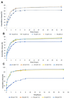
The cumulative releasing property of biomimetic thymine dimers 4a-d (A) and photo-adducts 6a-d (B) and 8a-d (C).
Figure 5
Figure 5
Panel (A): Binding mode of compound 8b. H-bond interactions are shown with continuous blue lines, and salt bridges are reported as dashed magenta lines. Panel (B): Binding mode of compound 8a. H-bond interactions are shown with continuous blue lines, and pi–cation interactions are reported as dashed red lines. Panel (C): Binding mode of compound 4b. H-bond interactions are shown with continuous blue lines. Panel (D): Binding mode of compound 4a. H-bond interactions are shown with continuous blue lines, hydrophobic interactions with dashed green lines, and pi-stacking interactions with dashed orange lines.

References

    1. Shen Y., Stanislauskas M., Li G., Zheng D., Liu L. Epigenetic and genetic dissections of UV-induced global gene dysregulation in skin cells through multi-omics analyses. Sci. Rep. 2017;7:42646. doi: 10.1038/srep42646. - DOI - PMC - PubMed
    1. Ravanat J.L., Douki T., Cadet J. Direct and indirect effects of UV radiation on DNA and its components. J. Photochem. Photobiol. B. 2001;63:88–102. doi: 10.1016/S1011-1344(01)00206-8. - DOI - PubMed
    1. Cadet J., Mouret S., Ravanat J.L., Douki T. Photoinduced damage to cellular DNA: Direct and photosensitized reactions. Photochem. Photobiol. 2012;88:1048–1065. doi: 10.1111/j.1751-1097.2012.01200.x. - DOI - PubMed
    1. Saha L.K., Wakasugi M., Akter S., Akter S., Prasad R., Wilson S.H., Shimizu N., Sasanuma H., Huang S.N., Agama K., et al. Topoisomerase I-driven repair of UV-induced damage in NER-deficient cells. Proc. Natl. Acad. Sci. USA. 2020;117:14412–14420. doi: 10.1073/pnas.1920165117. - DOI - PMC - PubMed
    1. Cadet J., Wagner J.R. DNA base damage by reactive oxygen species, oxidizing agents, and UV radiation. Cold Spring Harb. Perspect. Biol. 2013;5:a012559. doi: 10.1101/cshperspect.a012559. - DOI - PMC - PubMed

MeSH terms