. 2022 Jan 14;23(2):917.
doi: 10.3390/ijms23020917.

Astaxanthin Exerts Anabolic Effects via Pleiotropic Modulation of the Excitable Tissue

Affiliations

Astaxanthin Exerts Anabolic Effects via Pleiotropic Modulation of the Excitable Tissue

Mónika Gönczi et al. Int J Mol Sci. .

Abstract

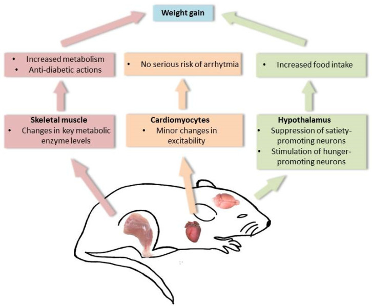
Astaxanthin is a lipid-soluble carotenoid influencing lipid metabolism, body weight, and insulin sensitivity. We provide a systematic analysis of acute and chronic effects of astaxanthin on different organs. Changes by chronic astaxanthin feeding were analyzed on general metabolism, expression of regulatory proteins in the skeletal muscle, as well as changes of excitation and synaptic activity in the hypothalamic arcuate nucleus of mice. Acute responses were also tested on canine cardiac muscle and different neuronal populations of the hypothalamic arcuate nucleus in mice. Dietary astaxanthin significantly increased food intake. It also increased protein levels affecting glucose metabolism and fatty acid biosynthesis in skeletal muscle. Inhibitory inputs innervating neurons of the arcuate nucleus regulating metabolism and food intake were strengthened by both acute and chronic astaxanthin treatment. Astaxanthin moderately shortened cardiac action potentials, depressed their plateau potential, and reduced the maximal rate of depolarization. Based on its complex actions on metabolism and food intake, our data support the previous findings that astaxanthin is suitable for supplementing the diet of patients with disturbances in energy homeostasis.

Keywords: arcuate nucleus; astaxanthin; cardiac action potential; excitability; food intake; gene expression; inhibitory postsynaptic current; metabolism; skeletal muscle.


Conflict of interest statement

The authors declare no conflict of interest.

Figures

Figure 1
Figure 1
Metabolic effects of chronic astaxanthin feeding. (A,B) Actions on O2 consumption without (A) and with normalization on body weight (B). Gray column: control group before sham feeding (C); hollow column: control group after sham feeding (S); black column: AX fed group before feeding of astaxanthin-containing chow (C); red column: AX fed group after feeding of astaxanthin-containing chow (AX). All panels have the same arrangement as above; (C,D) Effects on CO2 consumption without (C) and with normalization on body weight (D); (E) Normalized actions on food intake; (F) Changes in body weight gain. * denotes significant differences at p < 0.05.
Figure 2
Figure 2
Effects of astaxanthin feeding on the levels of signaling proteins in biceps femoris and pectoral muscles. (A) Statistical analysis of changes in signaling protein expression of the biceps femoris muscle; (B) statistical analysis of changes in signaling protein expression of the pectoral muscles. Black columns (C): control, red columns (AX): astaxanthin feeding. Quantification of the results is reported as average ± SEM (n = 3–5); * p < 0.05, ** p < 0.01, *** p < 0.001.
Figure 3
Figure 3
Effects of astaxanthin feeding on the excitability and synaptic currents of neurons in the hypothalamic arcuate nucleus. (A,B) Representative voltage traces of randomly chosen neurons of the arcuate nucleus in the control group (A, black) and in the AX fed population (B, red) elicited with 120 pA depolarizing current injection. (CE) Statistical analysis of changes in input resistance (C), maximal action potential firing rate (D) and average action potential firing rate at different levels of depolarization (E). Black columns and graphs: control group (C), red columns, and graphs: astaxanthin fed population (AX). (F,G) Excitatory (black arrows) and inhibitory (gray arrows) postsynaptic currents (EPSCs and IPSCs, respectively) recorded from the control group (F, black) and from the AX fed group (G, red). (H,I) Statistical analysis of the EPSC frequency and amplitude (H) and the IPSC frequency and amplitude (I). Black columns: control group (C), red columns: astaxanthin fed population (AX). * p < 0.05, ** p < 0.01.
Figure 4
Figure 4
Changes by acute astaxanthin feeding on POMC-positive neurons of the arcuate nucleus. (A) Evaluation of the neuronal type. Upper panel: POMC-driven tdTomato expression in red. Middle panel: biocytin labeling (green). Lower panel: merged image. Images represent a single confocal z-stack with 1-µm thickness. Scale bar: 50 µm; (B) voltage traces from a POMC-positive neuron under control conditions (black) and with acute 2.5 µM AX treatment (red) elicited with −30 and +120 pA current injections; (C) comparison of the average firing rate with different depolarizing steps under control conditions and during AX application (red); (D) synaptic currents of a POMC-positive neuron under control conditions (above) and during AX treatment (below). Black arrows: EPSCs, gray arrows: IPSCs; (E) a representative trace of histogram analysis of the holding current under control (black) and in the presence of astaxanthin (red); (F) statistical comparison of the spontaneous fluctuation of the holding current (C, black) and the effect of astaxanthin on the holding current (AX, red; hollow circles: individual data, filled squares: average ± SEM); (G,H) statistical analysis of the IPSC (G) and EPSC (H) frequency and amplitude. Black columns: control conditions (C), red columns: astaxanthin treatment (AX).
Figure 5
Figure 5
Changes in the excitability of the synaptic currents of the GABAergic inhibitory neurons of the arcuate nucleus. (A,B) Representative average calcium imaging traces from four GABAergic somata under control conditions and with application of 2.5 µM AX. (C) Statistical comparison of the calcium transient frequency of the GABAergic somata in control and with AX (black, C: control, red, AX) in the neuronal groups where increase, no change or decrease was seen (from left to right, respectively). Increase was seen in 77.3%, no change was observed in 9.1% and decrease was detected in 13.6% of the neuronal somata; (D,E) spontaneous activity of GABAergic neurons in control (black) and with AX (red); (F) statistical comparison of the spontaneous and normalized firing rate under control conditions and with AX (black, C: control, red, AX); (G) statistical representation of EPSC frequency in control (black, C) and with astaxanthin (red, AX); (H) average frequency change (black square) and frequency changes of the individual cases (green: increase, red: decrease, yellow: no change). * p < 0.05, ** p < 0.01.
Figure 6
Figure 6
Effects of astaxanthin on the parameters of action potentials in canine left ventricular myocytes. (A) Representative superimposed action potentials recorded in control (black) and in the presence of 2.5 µM astaxanthin (red). (B) First time derivative of the action potential upstroke with the same color code. (CJ) Average results obtained from 7 cells in control and in the presence of astaxanthin (black, C: control; red, AX: astaxanthin; columns and bars: mean ± SEM; asterisks: significant differences are noted by * with p < 0.05).
Figure 7
Figure 7
Proposed mode of action of AX in mice. Summary of our findings on the responses by AX on skeletal muscle metabolism, cardiac excitability and hypothalamic neuronal activity that could lead potentially to weight gain. AX facilitates the increase in metabolism and exerts anti-diabetic actions on skeletal muscle, promotes food intake via actions on the hypothalamus, and exerts minor changes on cardiac excitability.

Similar articles

References

    1. Nakao R., Nelson O.L., Park J.S., Mathison B.D., Thompson P.A., Chew B.P. Effect of astaxanthin supplementation on inflammation and cardiac function in BALB/c mice. Anticancer Res. 2010;30:2721–2725. - PubMed
    1. Ambati R.R., Phang S.M., Ravi S., Aswathanarayana R.G. Astaxanthin: Sources, extraction, stability, biological activities and its commercial applications—A review. Mar. Drugs. 2014;12:128–152. doi: 10.3390/md12010128. - DOI - PMC - PubMed
    1. Baralic Andjelkovic M., Djordjevic B., Dikic N., Radivojevic N., Suzin-Zivkovic V., Radojevic-Skodric S., Pejic S. Effect of astaxanthin supplementation on salivary IgA, oxidative stress, and inflammation in young soccer players. Evid. -Based Complement. Altern. Med. 2015;2015:783761. doi: 10.1155/2015/783761. - DOI - PMC - PubMed
    1. Hu J., Nagarajan D., Zhang Q., Chang J.S., Lee D.J. Heterotrophic cultivation ofmicroalgae for pigment production: A review. Biotechnol. Adv. 2017;36:54–67. doi: 10.1016/j.biotechadv.2017.09.009. - DOI - PubMed
    1. Sztretye M., Dienes B., Gönczi M., Czirják T., Csernoch L., Dux L., Szentesi P., Keller-Pintér A. Astaxanthin: A potential mitochondrial-targeted antioxidant treatment in diseases and with aging. Oxid. Med. Cell. Longev. 2019;2019:3849692. doi: 10.1155/2019/3849692. - DOI - PMC - PubMed

LinkOut - more resources