Review
. 2022 Jan 16;23(2):963.
doi: 10.3390/ijms23020963.

Inflammation as A Precursor of Atherothrombosis, Diabetes and Early Vascular Aging

Affiliations
Review

Inflammation as A Precursor of Atherothrombosis, Diabetes and Early Vascular Aging

Elena Barbu et al. Int J Mol Sci. .

Abstract

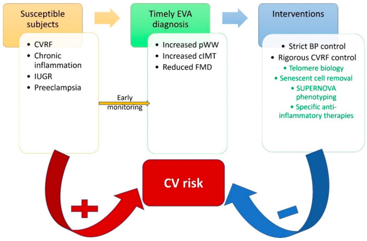
Vascular disease was for a long time considered a disease of the old age, but it is becoming increasingly clear that a cumulus of factors can cause early vascular aging (EVA). Inflammation plays a key role in vascular stiffening and also in other pathologies that induce vascular damage. There is a known and confirmed connection between inflammation and atherosclerosis. However, it has taken a long time to prove the beneficial effects of anti-inflammatory drugs on cardiovascular events. Diabetes can be both a product of inflammation and a cofactor implicated in the progression of vascular disease. When diabetes and inflammation are accompanied by obesity, this ominous trifecta leads to an increased incidence of atherothrombotic events. Research into earlier stages of vascular disease, and documentation of vulnerability to premature vascular disease, might be the key to success in preventing clinical events. Modulation of inflammation, combined with strict control of classical cardiovascular risk factors, seems to be the winning recipe. Identification of population subsets with a successful vascular aging (supernormal vascular aging-SUPERNOVA) pattern could also bring forth novel therapeutic interventions.

Keywords: COVID-19; NLPR3; SUPERNOVA; atherothrombosis; diabetes; inflammation; klotho; obesity; vascular senescence; visfatin.


Conflict of interest statement

The authors declare no conflict of interest.

Figures

Figure 1
Figure 1
The systemic effects of NLRP3 caspase-1-mediated interleukin activation. Interleukin-6 generated by the potent stimulus of IL-1b leads to secretion of acute-phase proteins in the liver, induces endothelial dysfunction and activates smooth muscle cells in arterial media. IL—interleukin; ox-LDL—oxidated LDL molecule; CRP—C-reactive protein; PAI-1—plasminogen activator inhibitor-1. (Reproduced with permission, [31]).
Figure 2
Figure 2
The molecular structure of the inflammasome. This is a large complex multimeric protein consisting of oligomerized NLRP. This consists of a central NOD immune receptor, a leucine-rich repeat (LRR) domain acting as an activation sensor and an effector domain, either a pyrin-containing (PYD) or a caspase activation and recruiting domain—CARD. When activated, the effector domain of NLRP interacts with an adaptor protein (ASC) via a CARD molecule and activates pro-caspase1 into mature caspase1. Pro-caspase1 also has a CARD molecule allowing interaction with the inflammasome. (Reproduced with permission, [31]).
Figure 3
Figure 3
The two main steps for full inflammasome activation. Initiation of the process is priming, which leads to increased expression of NLRP3 and pro-IL-1b via nuclear factor-kappa B. This acts as a second messenger for toll-like receptor activation via DAMPs or PAMPs; an alternative pathway is NF-kB stimulation by TNF receptor and TNF α. Triggering or full activation of the inflammasome is the final oligomerization of NLRP3 with procaspase-1 cleaving properties. It can be induced by intracellular signaling due to increased ROS generation by dysfunctional mitochondria, potassium efflux or calcium influx, or lysosomal lysis after LDL crystal endocytosis. (Reproduced with permission, [31]).
Figure 4
Figure 4
Protective vascular effects of Klotho proteins and adiponectin vs deleterious effects of NLRP and visfatin.
Figure 5
Figure 5
Proposed cardiovascular risk reduction strategy through identification of susceptible individuals, early diagnostic protocols and timely interventions. BP—blood pressure; cIMT—carotid intima-media thickness; CVRF—cardiovascular risk factors; FMD—flow-mediated dilation; IUGR—intrauterine growth restriction; pWW—pulse wave velocity. In green, promising future interventions.

References

    1. Damaskos C., Garmpis N., Kollia P., Mitsiopoulos G., Barlampa D., Drosos A., Patsouras A., Gravvanis N., Antoniou V., Litos A., et al. Assessing Cardiovascular Risk in Patients with Diabetes: An Update. Curr. Cardiol. Rev. 2019;16:266–274. doi: 10.2174/1573403X15666191111123622. - DOI - PMC - PubMed
    1. Glovaci D., Fan W., Wong N.D. Epidemiology of Diabetes Mellitus and Cardiovascular Disease. Curr. Cardiol. Rep. 2019;21:1–8. doi: 10.1007/s11886-019-1107-y. - DOI - PubMed
    1. Henning R.J. Type-2 diabetes mellitus and cardiovascular disease. Future Cardiol. 2018;14:491–509. doi: 10.2217/fca-2018-0045. - DOI - PubMed
    1. Shovman O., Levy Y., Gilburd B., Shoenfeld Y. Antiinflammatory and immunomodulatory properties of statins. Immunol. Res. 2002;25:271–285. doi: 10.1385/IR:25:3:271. - DOI - PubMed
    1. Cortese F., Gesualdo M., Cortese A., Carbonara S., Devito F., Zito A., Ricci G., Scicchitano P., Ciccone M.M. Rosuvastatin: Beyond the cholesterol-lowering effect. Pharmacol. Res. 2016;107:1–18. doi: 10.1016/j.phrs.2016.02.012. - DOI - PubMed