. 2021 Dec 24;13(1):25.
doi: 10.3390/insects13010025.

Anatomy of the Nervous System in Chelifer cancroides (Arachnida: Pseudoscorpiones) with a Distinct Sensory Pathway Associated with the Pedipalps

Affiliations

Anatomy of the Nervous System in Chelifer cancroides (Arachnida: Pseudoscorpiones) with a Distinct Sensory Pathway Associated with the Pedipalps

Torben Stemme et al. Insects. .

Abstract

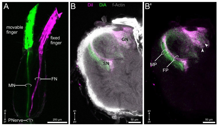
Many arachnid taxa have evolved unique, highly specialized sensory structures such as antenniform legs in Amblypygi (whip spiders), for instance, or mesosomal pectines in scorpions. Knowledge of the neuroanatomy as well as functional aspects of these sensory organs is rather scarce, especially in comparison to other arthropod clades. In pseudoscorpions, no special sensory structures have been discovered so far. Nevertheless, these animals possess dominant, multifunctional pedipalps, which are good candidates for being the primary sensory appendages. However, only little is known about the anatomy of the nervous system and the projection pattern of pedipalpal afferents in this taxon. By using immunofluorescent labeling of neuronal structures as well as lipophilic dye labeling of pedipalpal pathways, we identified the arcuate body, as well as a comparatively small mushroom body, the latter showing some similarities to that of Solifugae (sun spiders and camel spiders). Furthermore, afferents from the pedipalps terminate in a glomerular and a layered neuropil. Due to the innervation pattern and structural appearance, we conclude that these neuropils are the first integration centers of the chemosensory and mechanosensory afferents. Within Arthropoda, but also other invertebrates or even vertebrates, sensory structures show rather similar neuronal arrangement. Thus, these similarities in the sensory systems of different evolutionary origin have to be interpreted as functional prerequisites of the respective modality.

Keywords: Chelicerata; afferents; brain; chemosensation; chemotopy; immunofluorescence; mechanosensation; morphology; olfaction; somatotopy.


Conflict of interest statement

The authors declare no conflict of interest. The funders had no role in the design of the study; in the collection, analyses, or interpretation of data; in the writing of the manuscript, or in the decision to publish the results.

Figures

Figure 1
Figure 1
General morphology, the sensory equipment, and neuronal innervation of the pedipalps of Chelifer cancroides. (A) Dorsal view of the animal, showing the four pairs of walking legs, the chelicera, and the large pedipalps, consisting of the trochanter, femur, patella, and the chela (tibia + tarsus). All appendages are associated with the prosoma (PR). The opisthosoma (OP) does not possess any appendages. (B) Overview of the chelal surface stained with Congo red (gray). The chela possesses two fingers, a dorsal fixed finger, and a ventral movable finger (joint indicated by arrowhead). (C) Detail of the movable finger, showing the sensory equipment with different types of sensilla, e.g., trichobothria (arrows), shorter hair-sensilla (double arrowheads), and club-shaped sensilla (arrowheads). The chelal teeth (CT) and poison tooth (PT) are indicated. (D) Overview of the innervation of the chela based on lipophilic dye injection (DiI, yellow). Within the chelal hand, two main fibers can be distinguished, each innervating one of the fingers (FN: fixed finger nerve; MN: movable finger nerve). Shortly after entering the fingers, these nerves split into two branches (veFN, doFN: ventral and dorsal nerve of fixed finger; veMN, doMN: ventral and dorsal nerve of movable finger). (E,E’) Details of the innervation of the chelal hand. From the main nerve branches (FN shown), various branches split of and send fine branches (double arrowheads in (E)) toward sensory structures at the cuticle surface (arrows in (E)). Along these fine fibers, swellings of cell bodies can be observed (arrowheads in (E,E’)). (F,F’) Details of the innervation of the fixed finger. From the dorsal and ventral nerve branches within the fingers, numerous fibers branch off and innervate sensory structures (double arrowheads in (F,F’)). Again, swellings consisting of cell bodies are present (arrowhead in (F’)). Other abbreviations: di: distal; do: dorsal; pr: proximal; ve: ventral.
Figure 2
Figure 2
General morphology of the central nervous system of Chelifer cancroides, revealed by fluorescent labeling of synaptic regions (green), f-actin (gray), and cell bodies (blue). (A) Position of the synganglion within the prosoma from a dorsal view. The dashed line indicates the outline of the synganglion. (B) From a sagittal view at the midline level, the synganglion is positioned at the dorsal margin of the prosoma. The soma cortex (CO) surrounds the neuropilar regions. The synganglion is divided into a supraesophageal brain (BR) and the subesophageal ganglion (SOG). Dashed line indicates the esophagus. (C) Sagittal slice of the synganglion at a more lateral level compared to (B). Ventrally, the four walking leg neuromeres (WLN1–4) can be recognized. Anterior to the WLN lies the large pedipalpal neuromere (PN). Dorsally to the PN, the cheliceral neuromere (CN) can be distinguished. The main part of the brain consists of the protocerebrum (PC), including the dorso-posterior arcuate body (AB). (D) Dorsal section of the synganglion at the level of the first walking leg neuromere (WLN1). A thick fiber tract (WLT1) connects the lateral parts of the WLN1 with medial areas of the synganglion. The WLT1 splits into three branches, a ventral (VHT1), an intermediate (IHT1) and a dorsal horizontal tract (DHT1). At least the IHT1 and the DHT1 seem to contain contralateral projections (double arrowheads). (E) Horizontal section of the synganglion at a ventral level. The ventral horizontal tracts of the walking legs (VHTs) are connected to a ventral longitudinal tract near the midline (VLT). Arrowheads point on two commissures formed by the bilateral symmetric VLTs. (F) At a more dorsal level, a thick, more lateral longitudinal tract (LLT) appears. Between these bilateral symmetric tracts, paired (arrows) and unpaired midline neuropils (double arrowhead) can be distinguished. (G) At an even more dorsal level, the dorsal horizontal tracts of the walking legs split up in several dorsal longitudinal tracts (arrows). Note the fused mass of opisthosomal ganglia (OPN), between the fourth walking leg neuromeres (WLN4). Other abbreviations: an: anterior; CC: chelicera; do: dorsal; po: posterior; PP: pedipalp; ve: ventral; WL1–4: walking legs 1–4.
Figure 3
Figure 3
Neuroanatomical details of the protocerebrum in Chelifer cancroides, revealed by fluorescent labeling of synaptic regions (green), f-actin (gray), and cell bodies (blue). (A,B) Frontal (A) and sagittal view on the protocerebrum. The arcuate body (AB) is located at a postero-dorsal position and consists of a dorsal (doAB) and a ventral layer (veAB). (C) In horizontal view, the arcuate body is built up of several palisade-like subunits, which are separated by fiber tracts (double arrowheads in (C) and (D)). (D) Distinct fibers (arrowheads) connect the arcuate body and contralateral protocerebral neuropils. (EG) Ventral to dorsal series of the protocerebrum, showing the components of the mushroom bodies. Two fiber tracts (arrowheads in (E)) emerge from an antero-lateral neuropil (arrow in (E)). More ventrally both fiber tracts (arrowheads in (F)) fuse and project further posteriorly. At a dorsal level, the fused main tract (white arrowhead in (G)) terminates in a postero-medial lobe (double arrowhead in (G)) close to the midline. A lateral tract leaves the main tract (black arrowhead in (G)). It seems, that the main tract projects further posteriorly (arrow in (G)) and terminates in a second lobe (asterisk in (G)) in close vicinity to the arcuate body. Other abbreviations: an: anterior; do: dorsal; po: posterior; ve: ventral.
Figure 4
Figure 4
Position and morphology of glomerular structures in the pedipalpal neuropil, revealed by fluorescent labeling of synaptic regions (green), f-actin (gray), and cell bodies (blue). (A) In the most dorsal horizontal section (in relation of the subesophageal ganglion) of the synganglion, a conspicuous, drop-shaped neuropil (GN) consisting of glomerular subunits can be observed in antero-medial aspects of the pedipalpal neuromere. Note the commissure in posterior region (arrowhead). (B,C) In the sagittal view, the glomerular neuropil (GN) spans spherically around the esophagus, with a fraction lying dorsally, the other part lying ventrally with respect to the esophagus (dashed lines). Asterisks indicate glomeruli. (D) In the dorsal view, the arrangement of the glomeruli (asterisks) gives the impression of separated subcompartments (indicated by dashed lines). (E) The glomerular neuropil is penetrated by fibers (arrowheads) from the antero-lateral direction, innervating the glomeruli (asterisks). (F) Besides the glomerular neuropil, another distinct neuropil is found postero-laterally, having a stratified appearance of parallel fibers (arrowheads). (GI) Frontal views on the glomerular neuropil (highlighted by dashed lines), showing a spheric to drop-shaped appearance. The glomerular neuropil is connected to dorsal, protocerebral regions by two distinct tracts (rings in (H)). Ventrally, fibers connect the glomerular neuropil to the ventral soma cortex (rings in (I)). Other abbreviations: an: anterior; CN: cheliceral neuropil; do: dorsal; PC: protocerebrum; po: posterior; ve: ventral.
Figure 5
Figure 5
Innervation of the pedipalpal neuropil revealed by lipophilic dye labeling (DiI, yellow), injected at the distal part of the patella, combined with fluorescent labeling of the synaptic regions (green) and cell bodies (blue). (A) Overview of a dye injected pedipalp, visualizing the projection pattern of the pedipalpal nerve (PNerve). (B,C) Maximum projections of two consecutive horizontal sections, from ventral (B) to dorsal (C). Dye-labelled fibers enter the glomerular (GN) and the stratified neuropil (SN). Further, a small neuropil between GN and SN is labelled (arrowheads; accessory neuropil: AN). (D) Detail of the pedipalpal neuropil (PN), horizontal view. The glomeruli (asterisks) are clearly labelled. Furthermore, the accessory neuropil (AN) and the medial portion of the stratified neuropil (SN) are marked. Note that no labeling is evident in the contralateral GN (dashed line). (E,E’,E”) Horizontal section of the pedipalpal neuropil labelled against synapsin (E) and by lipophilic dye (E’); both channels merged in (E”). Glomeruli are indicated by asterisks. Note the layered innervation of the stratified neuropil, indicated by rings in (E”). The AN seems to receive input from anterior fibers associated with the SN (arrowheads in (E’,E”)). Other abbreviations: an: anterior; di: distal; po: posterior; pr: proximal; WLN1–4: walking leg neuromeres 1–4.
Figure 6
Figure 6
Differential labeling of movable and fixed finger nerves with lipophilic dyes (DiI: magenta; DiA: green), combined with f-actin labeling (gray). (A) Overview of dye-injected fingers, visualizing the projection pattern of the fixed finger (FN) and movable finger nerve (MN). At the chela–patella joint, both fibers fuse (pedipalpal nerve: PNerve). (B,B’) Horizontal view, showing the innervation pattern of the glomerular neuropil (GN) and the stratified neuropil (SN). While the GN is innervated homogenously (arrows point to selected glomeruli), the stratified neuropil shows a distinct innervation pattern by fibers associated with the movable finger (movable finger portion: MP) and fixed finger (fixed finger portion: FP). Other abbreviations: an: anterior; di: distal; po: posterior; pr: proximal.
Figure 7
Figure 7
Schematic summary of the projection pattern of the sensory structures associated with the pedipalpal chelae of Chelifer cancroides. Differential labeling of afferents of the fixed (FN: fixed finger nerve, magenta) and movable finger (MN: movable finger nerve, green) innervate two neuropils in the pedipalpal neuromere within the central nervous system (CNS): The glomerular neuropil (GN) is innervated rather homogenously by projections from both fingers, while the stratified neuropil (SN) shows a clear separation in an anterior portion innervated by afferents from the fixed finger and a posterior portion innervated by fibers associated with the movable finger. Other abbreviations: AN: accessory neuropil; PNerve: pedipalpal nerve.

References

    1. Harvey M.S. The smaller arachnid orders: Diversity, descriptions and distributions from Linnaeus to the present (1758 to 2007) Zootaxa. 2007;1668:363–380. doi: 10.11646/zootaxa.1668.1.19. - DOI
    1. Levi H.W. Observation on two species of pseudoscorpions. Can. Entomol. 1953;85:55–62. doi: 10.4039/Ent8555-2. - DOI
    1. Weygoldt P. Biology of Pseudoscorpions. 1st ed. Harvard University Press; Harvard, UK: 1969. pp. 1–159.
    1. Brach V. Social behavior in the pseudoscorpion Paratemnus elongatus (Banks) (Pseudoscorpionida, Atemnidae) Insect Soc. 1978;25:3–11. doi: 10.1007/BF02224481. - DOI
    1. Zeh D.W. Life history consequences of sexual dimorphism in a chernetid pseudoscorpion. Ecology. 1987;68:1495–1501. doi: 10.2307/1939233. - DOI

LinkOut - more resources