. 2022 Jan 4;14(1):113.
doi: 10.3390/pharmaceutics14010113.

Elovanoids Counteract Inflammatory Signaling, Autophagy, Endoplasmic Reticulum Stress, and Senescence Gene Programming in Human Nasal Epithelial Cells Exposed to Allergens

Affiliations

Elovanoids Counteract Inflammatory Signaling, Autophagy, Endoplasmic Reticulum Stress, and Senescence Gene Programming in Human Nasal Epithelial Cells Exposed to Allergens

Alfredo Resano et al. Pharmaceutics. .

Abstract

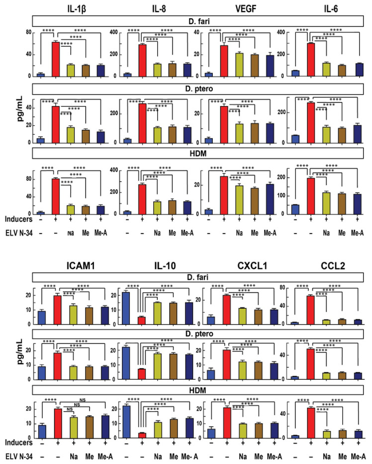
To contribute to further understanding the cellular and molecular complexities of inflammatory-immune responses in allergic disorders, we have tested the pro-homeostatic elovanoids (ELV) in human nasal epithelial cells (HNEpC) in culture challenged by several allergens. ELV are novel bioactive lipid mediators synthesized from the omega-3 very-long-chain polyunsaturated fatty acids (VLC-PUFA,n-3). We ask if: (a) several critical signaling events that sustain the integrity of the human nasal epithelium and other organ barriers are perturbed by house dust mites (HDM) and other allergens, and (b) if ELV would participate in beneficially modulating these events. HDM is a prevalent indoor allergen that frequently causes allergic respiratory diseases, including allergic rhinitis and allergic asthma, in HDM-sensitized individuals. Our study used HNEpC as an in vitro model to study the effects of ELV in counteracting HDM sensitization resulting in inflammation, endoplasmic reticulum (ER) stress, autophagy, and senescence. HNEpC were challenged with the following allergy inducers: LPS, poly(I:C), or Dermatophagoides farinae plus Dermatophagoides pteronyssinus extract (HDM) (30 µg/mL), with either phosphate-buffered saline (PBS) (vehicle) or ELVN-34 (500 nM). Results show that ELVN-34 promotes cell viability and reduces cytotoxicity upon HDM sensitization of HNEpC. This lipid mediator remarkably reduces the abundance of pro-inflammatory cytokines and chemokines IL-1β, IL-8, VEGF, IL-6, CXCL1, CCL2, and cell adhesion molecule ICAM1 and restores the levels of the pleiotropic anti-inflammatory IL-10. ELVN-34 also lessens the expression of senescence gene programming as well as of gene transcription engaged in pro-inflammatory responses. Our data also uncovered that HDM triggered the expression of key genes that drive autophagy, unfolded protein response (UPR), and matrix metalloproteinases (MMP). ELVN-34 has been shown to counteract these effects effectively. Together, our data reveal a novel, pro-homeostatic, cell-protective lipid-signaling mechanism in HNEpC as potential therapeutic targets for allergies.

Keywords: allergy; anti-inflammatory; elovanoids; house dust mite; inflammation; lipid mediators.


Conflict of interest statement

The authors declare no conflict of interest. Diater Laboratorio had no role in the design of the study; in the collection, analyses, or interpretation of data; in the writing of the manuscript, and in the decision to publish the results.

Figures

Figure 1
Figure 1
Experimental design. Human nasal epithelial cells (HNEpC) in primary culture challenged by allergy inducers—LPS, poly (I:C), D. fari, D. ptero, or a combination of house dust mite extracts (HDM) (D. farinae + D. pteronyssinus) (30 µg/mL) were analyzed to determine the protective effect of elovanoids (ELV)—ELVN-34-Na, 34-ME, or 34-ME-A (500 nM).
Figure 2
Figure 2
ELVN-34 protect HNEpC from cytotoxicity induced by HDM. Cell viability assay using Presto Blue HS kit (left panel) indicated that HNEpC treated with ELVN-34 (500 nM) were protected from cytotoxicity and cell death induced by Dermatophagoides farinae, Dermatophagoides pteronyssinus, and (D. farinae + D. pteronyssinus) (HDM), or LPS or poly(I:C) (30 μg/mL). Cytotoxicity assay using CyQuant LDH (right panel) shows that ELVN-34 (500 nM) elicit potent cytoprotection to HNEpC when challenged by the inducers. The results showed the averages of three independent experiments (**** p < 0.0001).
Figure 3
Figure 3
ELVN-34 reduce the expression of pro-inflammatory cytokines, chemokines and cell adhesion molecule in HNEpC challenged with LPS or poly(I:C). HNEpC challenged with LPS or poly(I:C) (30 μg/mL) display enhanced production of pro-inflammatory cytokines and chemokines IL-6, IL-1β, IL-8/CXCL8, CCL2/MCP-1, CXCL1/KC/GRO, VEGF, and cell adhesion molecule ICAM1(CD54) compared to non-treated cells. This increase in the production of pro-inflammatory molecules is abrogated by the addition of ELVN-34 (500 nM) 30 min post-challenge. LPS and poly(I:C) induce a reduction of anti-inflammatory IL-10 expression in HNEpC that is restored to normal levels after treatment with ELVN-34 (500 nM). The results showed the averages of three independent experiments. (**** p < 0.0001, NS—not significant).
Figure 4
Figure 4
ELVN-34 reduce the expression of pro-inflammatory cytokines, chemokines, and cell adhesion molecule in HNEpC challenged with D. farinae, D. pteronyssinus, and HDM (D. fari + D. ptero) (30 μg/mL). ELVN-34 reduce the expression of pro-inflammatory cytokines, chemokines, and cell adhesion molecule ICAM1 in HNEpC exposed to HDM inducers. A remarkable decrease was observed in the release of anti-inflammatory cytokine IL-10 in HNEpC challenged with the HDM stressors (D. farinae, D. pteronyssinus, and HDM) (30 μg/mL). This decreased production of anti-inflammatory cytokine is reversed by the addition of ELVN-34 (500 nM), 30 min post-challenge with the stressor. The results showed the averages of three independent experiments. (**** p < 0.0001).
Figure 5
Figure 5
ELVN-34 protect HNEpC from the senescence-associated secretory phenotype (SASP response). Representative images of HNEpC stained with Hoechst 33342 and spider β-galactosidase—(A) untreated (control), (B) challenged with HDM (30 μg/mL), and (C) HNEpC treated with HDM and ELVN-34:6 Na (500 nM). (D) Quantitative assessment of increased SASP response (measured by the spider β-galactosidase staining) upon HDM exposure (30 μg/mL). ELVN-34:6 Na (500 nM) treatment reduces the number of beta-gal-positive HNEpC induced by HDM. The results showed the averages of three independent experiments. (* p < 0.05, NS—not significant).
Figure 6
Figure 6
HDM triggered multiple signaling in HNEpC. HNEpC challenged with HDM (D. farinae + D. pteronyssinus) (30 μg/mL) induces the expression of several genes related with autophagy (ATG3, ATG5, ATG7, BECLIN-1, and P62), unfolded protein response (UPR) (ATF6, CHOP, and IRE1), and matrix metalloproteinases (MMPs) (MMP8, MMP2, MMP9, MMP3, MMP12, TIMP1, and TIMP2). HDM stressors also induce the expression of senescence (P21, P16, P27, and P53) and inflammation genes (IL-1α, IL-6, and IL-1β) on HNEpC. The treatment with ELVN-34 Na (500 nM) reduces the expression of autophagy, UPR, MMP, senescence (except P53), and inflammation genes induced by HDM extracts. The results showed the averages of three independent experiments. (**** p < 0.0001, NS—not significant).

References

    1. Calderón M.A., Linneberg A., Kleine-Tebbe J., De Blay F., Hernandez Fernandez de Rojas D., Virchow J.C., Demoly P. Respiratory allergy caused by house dust mites: What do we really know? J. Allergy Clin. Immunol. 2015;136:38–48. doi: 10.1016/j.jaci.2014.10.012. - DOI - PubMed
    1. Dumez M.-E., Herman J., Campizi V., Galleni M., Jacquet A., Chevignà A. Orchestration of an Uncommon Maturation Cascade of the House Dust Mite Protease Allergen Quartet. Front. Immunol. 2014;5:138. doi: 10.3389/fimmu.2014.00138. - DOI - PMC - PubMed
    1. Foray A.-P., Dietrich C., Pecquet C., Machavoine F., Chatenoud L., Leite-de-Moraes M. IL-4 and IL-17 Are Required for House Dust Mite-Driven Airway Hyperresponsiveness in Autoimmune Diabetes-Prone Non-Obese Diabetic Mice. Front. Immunol. 2021;11:595003. doi: 10.3389/fimmu.2020.595003. - DOI - PMC - PubMed
    1. Gandhi V.D., Davidson C., Asaduzzaman M., Nahirney D., Vliagoftis H. House Dust Mite Interactions with Airway Epithelium: Role in Allergic Airway Inflammation. Curr. Allergy Asthma Rep. 2013;13:262–270. doi: 10.1007/s11882-013-0349-9. - DOI - PubMed
    1. Gregory L.G., Lloyd C.M. Orchestrating house dust mite-associated allergy in the lung. Trends Immunol. 2011;32:402–411. doi: 10.1016/j.it.2011.06.006. - DOI - PMC - PubMed

LinkOut - more resources