. 2022 Jan 5;14(1):111.
doi: 10.3390/pharmaceutics14010111.

Tailoring of Novel Azithromycin-Loaded Zinc Oxide Nanoparticles for Wound Healing

Affiliations

Tailoring of Novel Azithromycin-Loaded Zinc Oxide Nanoparticles for Wound Healing

Mohammed S Saddik et al. Pharmaceutics. .

Abstract

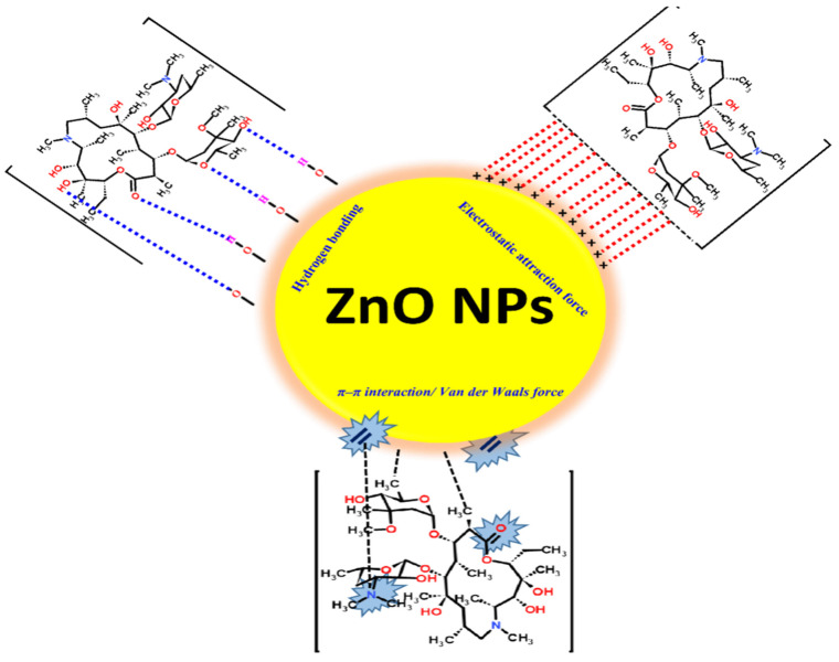
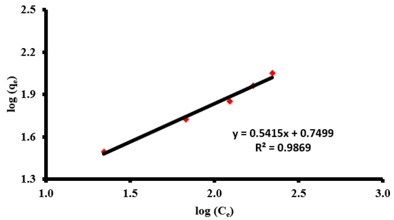
Skin is the largest mechanical barrier against invading pathogens. Following skin injury, the healing process immediately starts to regenerate the damaged tissues and to avoid complications that usually include colonization by pathogenic bacteria, leading to fever and sepsis, which further impairs and complicates the healing process. So, there is an urgent need to develop a novel pharmaceutical material that promotes the healing of infected wounds. The present work aimed to prepare and evaluate the efficacy of novel azithromycin-loaded zinc oxide nanoparticles (AZM-ZnONPs) in the treatment of infected wounds. The Box-Behnken design and response surface methodology were used to evaluate loading efficiency and release characteristics of the prepared NPs. The minimum inhibitory concentration (MIC) of the formulations was determined against Staphylococcus aureus and Escherichia coli. Moreover, the anti-bacterial and wound-healing activities of the AZM-loaded ZnONPs impregnated into hydroxyl propyl methylcellulose (HPMC) gel were evaluated in an excisional wound model in rats. The prepared ZnONPs were loaded with AZM by adsorption. The prepared ZnONPs were fully characterized by XRD, EDAX, SEM, TEM, and FT-IR analysis. Particle size distribution for the prepared ZnO and AZM-ZnONPs were determined and found to be 34 and 39 nm, respectively. The mechanism by which AZM adsorbed on the surface of ZnONPs was the best fit by the Freundlich model with a maximum load capacity of 160.4 mg/g. Anti-microbial studies showed that AZM-ZnONPs were more effective than other controls. Using an experimental infection model in rats, AZM-ZnONPs impregnated into HPMC gel enhanced bacterial clearance and epidermal regeneration, and stimulated tissue formation. In conclusion, AZM -loaded ZnONPs are a promising platform for effective and rapid healing of infected wounds.

Keywords: azithromycin; metal nanoparticles; wound healing; zinc oxide nanoparticles.


Conflict of interest statement

The authors declare no conflict of interest.

Figures

Scheme 1
Scheme 1
Synthesis pathway for the investigated ZnONPs.
Figure 1
Figure 1
(A) FT-IR spectra of ZnONPs, AZM, and AZM-ZnO nanoparticles; (B) typical XRD patterns of the prepared ZnO nanoparticles (note: The numbers above the peaks correspond to the miller index (hkl) values of hexagonal ZnO); (C) EDXS spectra of the investigated ZnONPs.
Figure 2
Figure 2
SEM image of (A) ZnONPs and (B) AZM-ZnONPs; TEM images of (C) ZnONPs and (E) AZM-ZnONPs and their particle size distribution in (D,F), respectively.
Figure 3
Figure 3
Removal performance of the AZM at different concentrations via ZnONPs.
Scheme 2
Scheme 2
AZM-ZnONP binding mechanism probabilities.
Figure 4
Figure 4
Freundlich isotherm model of the adsorption of AZM via ZnONPs.
Figure 5
Figure 5
Surface plot effects of different formulation factors on LE%.
Figure 6
Figure 6
Surface plot effects of different formulation factors on release after 1 h.
Figure 7
Figure 7
Surface plot effects of different formulation factors on release after 3 h.
Figure 8
Figure 8
Azithromycin-loaded zinc oxide nanoparticles have superior anti-bacterial activities. (A) shows the mean inhibition zone diameters induced by azithromycin (AZM), zinc oxide (ZnO), and azithromycin-loaded zinc oxide nanoparticles (AZM-ZnONPs) in agar plates inoculated by MRSA and E. coli, and a representative figure of each treatment. (B) shows the minimum inhibitory concentrations (MIC) of AZM, ZnO, and AZM-loaded ZnONPs against MRSA and E. coli. Data are shown as mean ± standard deviation of 3 experiments.
Figure 9
Figure 9
In vivo analysis of the tested preparations. (A) Wounds of rats were inoculated with MRSA and treated with indicated preparations then photographed at 1, 3, and 7 days post infection to visualize the healing process. (B) Bacterial load burden was evaluated in the wounded skin at 1, 3, and 7 days post infection. The results are expressed as the means ± SD. CFU, colony forming units. (C) Effect of AZM, ZnO, and AZM-ZnONPs on the percentage of wound contraction was evaluated over a duration of 10 days after infection.
Figure 10
Figure 10
Hematoxylin-and-eosin-stained sections of skin from rats from all groups. (a) Negative control group, (b) untreated control group, (c) AZM-treated group, (d) zinc oxide-treated group, (e) AZM-ZnONP-treated group, and (f) mean epidermal thickness of skin in different groups. Data are expressed as mean ± SD. p-values were calculated with the Mann–Whitney test. ** p value < 0.005. Insets represent magnified sections. Epidermis (E), dermis (D), dermal–epidermal junction (arrowhead), and hair follicles (arrow).
Figure 11
Figure 11
Masson’s trichrome-stained sections of skin from rats from all groups. (a) Negative control group, (b) untreated control group, (c) AZM-treated group, (d) zinc oxide-treated group, (e) AZM-ZnONP-treated group, and (f) mean percentage of collagen fibers of dermis in different groups. Data are expressed as mean ± SD. p-values were calculated with the Mann–Whitney test. * p value < 0.05. ** p value < 0.005. Double arrows refer to collagen fibers.

References

    1. Velnar T., Bailey T., Smrkolj V. The wound healing process: An overview of the cellular and molecular mechanisms. J. Int. Med. Res. 2009;37:1528–1542. doi: 10.1177/147323000903700531. - DOI - PubMed
    1. WYNN M. The impact of infection on the four stages of acute wound healing: An overview. Wounds UK. 2021;17:26–32.
    1. Teaima M.H., Elasaly M.K., Omar S.A., El-Nabarawi M.A., Shoueir K.R. Wound healing activities of polyurethane modified chitosan nanofibers loaded with different concentrations of linezolid in an experimental model of diabetes. J. Drug Deliv. Sci. Technol. 2021 doi: 10.1016/j.jddst.2021.102982. - DOI
    1. Fathi H.A., Abdelkader A., AbdelKarim M.S., Abdelaziz A.A., El-Mokhtar M.A., Allam A., Fetih G., El Badry M., Elsabahy M. Electrospun vancomycin-loaded nanofibers for management of methicillin-resistant Staphylococcus aureus-induced skin infections. Int. J. Pharm. 2020;586:119620. doi: 10.1016/j.ijpharm.2020.119620. - DOI - PubMed
    1. Al-Hakkani M.F., Gouda G.A., Hassan S.H.A. A review of green methods for phytofabrication of hematite (α-Fe2O3) nanoparticles and their characterization, properties, and applications. Heliyon. 2021;7:e05806. doi: 10.1016/j.heliyon.2020.e05806. - DOI - PMC - PubMed

LinkOut - more resources