. 2022 Jan 7;14(1):143.
doi: 10.3390/pharmaceutics14010143.

Enzyme-Responsive Amphiphilic Peptide Nanoparticles for Biocompatible and Efficient Drug Delivery

Affiliations

Enzyme-Responsive Amphiphilic Peptide Nanoparticles for Biocompatible and Efficient Drug Delivery

Su Jeong Song et al. Pharmaceutics. .

Abstract

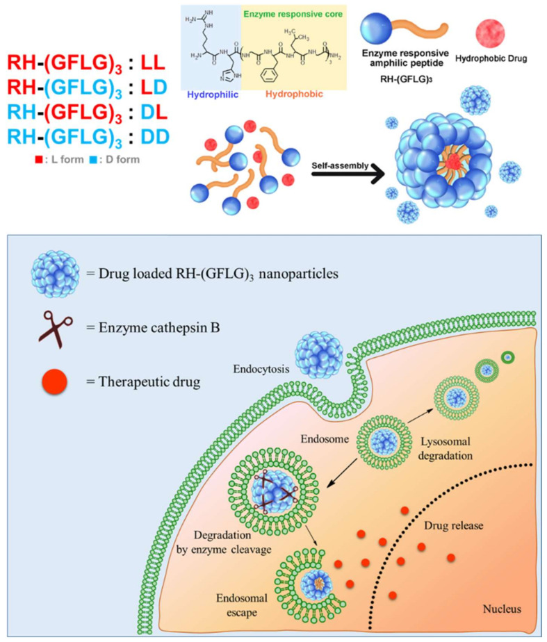
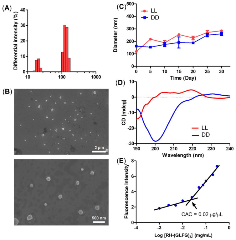
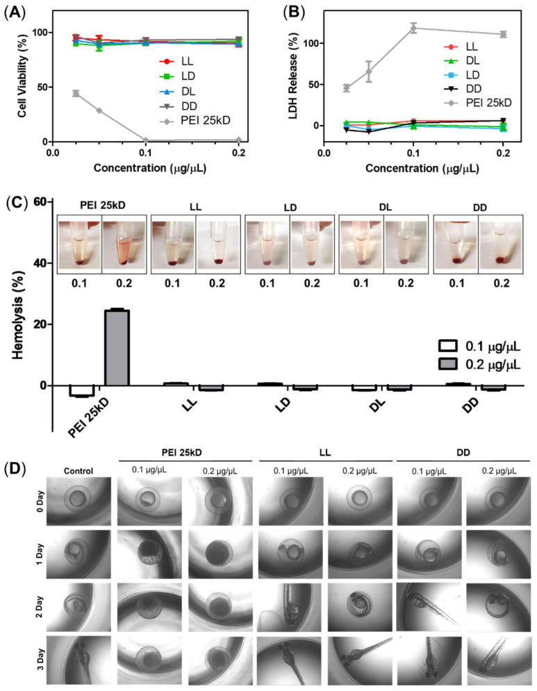
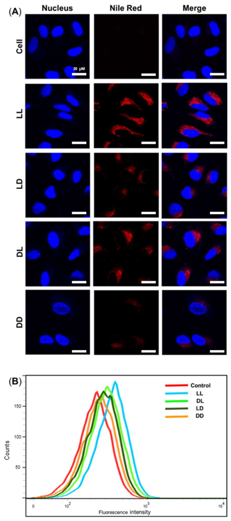
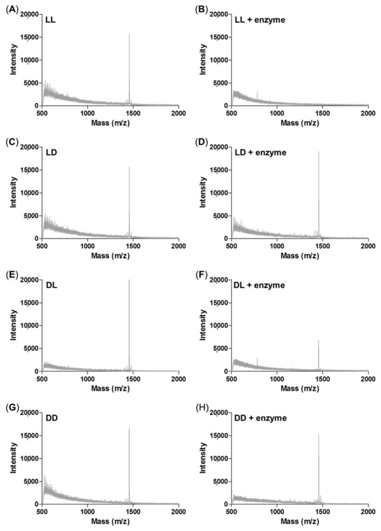
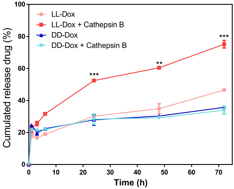
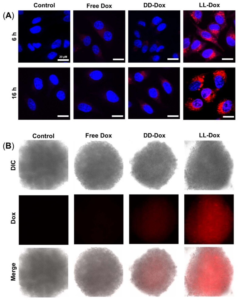
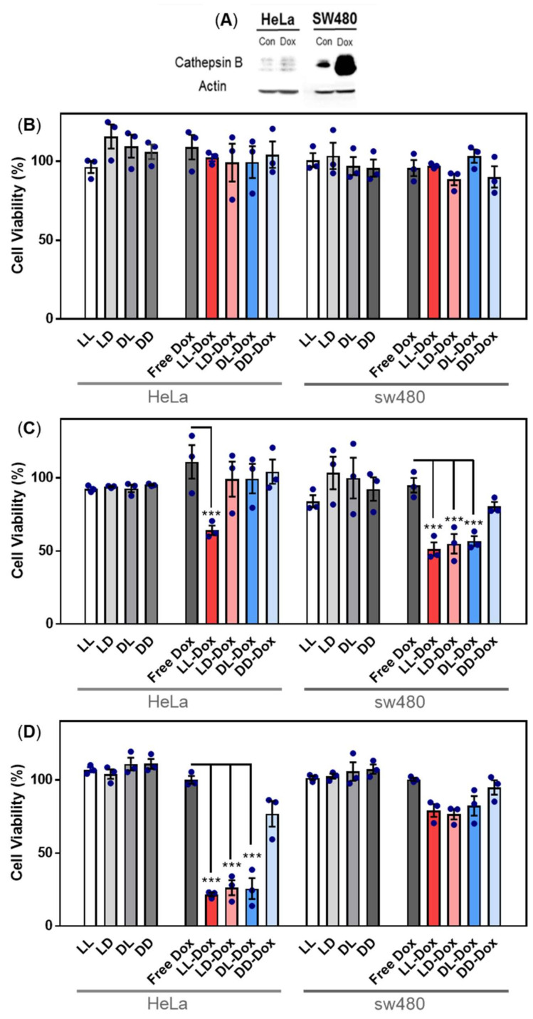
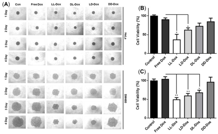
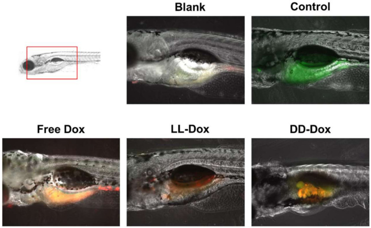
Self-assembled peptide nanostructures recently have gained much attention as drug delivery systems. As biomolecules, peptides have enhanced biocompatibility and biodegradability compared to polymer-based carriers. We introduce a peptide nanoparticle system containing arginine, histidine, and an enzyme-responsive core of repeating GLFG oligopeptides. GLFG oligopeptides exhibit specific sensitivity towards the enzyme cathepsin B that helps effective controlled release of cargo molecules in the cytoplasm. Arginine can induce cell penetration, and histidine facilitates lysosomal escape by its buffering capacity. Herein, we propose an enzyme-responsive amphiphilic peptide delivery system (Arg-His-(Gly-Phe-Lue-Gly)3, RH-(GFLG)3). The self-assembled RH-(GFLG)3 globular nanoparticle structure exhibited a positive charge and formulation stability for 35 days. Nile Red-tagged RH-(GFLG)3 nanoparticles showed good cellular uptake compared to the non-enzyme-responsive control groups with d-form peptides (LD (LRH-D(GFLG)3), DL (DRH-L(GFLG)3), and DD (DRH-D(GFLG)3). The RH-(GFLG)3 nanoparticles showed negligible cytotoxicity in HeLa cells and human RBCs. To determine the drug delivery efficacy, we introduced the anticancer drug doxorubicin (Dox) in the RH-(GFLG)3 nanoparticle system. LL-Dox exhibited formulation stability, maintaining the physical properties of the nanostructure, as well as a robust anticancer effect in HeLa cells compared to DD-Dox. These results indicate that the enzyme-sensitive RH-(GFLG)3 peptide nanoparticles are promising candidates as drug delivery carriers for biomedical applications.

Keywords: cytotoxicity; doxorubicin; drug delivery systems; peptide nanoparticle.


Conflict of interest statement

The authors declare no conflict of interest.

Figures

Figure 1
Figure 1
Schematic illustration of RH-(GFLG)3 nanoparticles as a drug carrier.
Figure 2
Figure 2
Physical characterization of the RH-(GFLG)3 nanoparticles. (A) Size distribution of LL nanoparticles. (B) Field emission-scanning electron microscopy image. (C) Size change of LL and DD nanoparticles for a storage time of 30 days. The size distribution was measured every 5 days. Values are reported as mean ± SEM (n = 3). (D) Circular dichroism (CD) spectrum of LL and DD nanoparticles. (E) Critical aggregation concentration (CAC) of LL nanoparticles.
Figure 3
Figure 3
Cytotoxicity of RH-(GFLG)3 nanoparticles. Cell viability using (A) MTT assay and (B) LDH assay. Concentrations of 0.2, 0.1, 0.5, and 0.25 µg/µL were tested for an incubation time of 24 h in HeLa cells. Values are reported as mean ± SEM (n = 3). (C) Hemolysis assay testing concentrations of 0.1 and 0.2 µg/µL using human blood cells. PEI 25kD was used as positive control. Values are reported as mean ± SEM (n = 3). (D) Zebrafish embryo test performed with nanoparticles at concentrations of 0.1 and 0.2 µg/µL. The incubation time was 3 days.
Figure 4
Figure 4
Cellular uptake assay of the Nile Red-tagged RH-(GFLG)3 nanoparticles. (A) Fluorescence images obtained via confocal microscopy of cell, LL, LD, DL, and DD following incubation for 6 h. Scale bar = 20 µm (blue = nucleus, red = RH-(GFLG)3 nanoparticles). (B) Fluorescence-activated cell sorting (FACS) analysis of cells incubated with control (empty peptide nanoparticles), LL, LD, DL, and DD during an incubation time of 16 h.
Figure 5
Figure 5
Enzyme sensitivity assay using MALDI-TOF mass spectrometry analysis. (A) LL peptide control; (B) LL peptide by cathepsin B treatment; (C) LD peptide control; (D) LD peptide by cathepsin B treatment; (E) DL peptide control; (F) DL peptide by cathepsin B treatment; (G) DD peptide control; (H) DD peptide by cathepsin B treatment.
Figure 6
Figure 6
In vitro drug release test of LL-Dox and DD-Dox by enzyme treatment. Tested incubation times were 0, 1, 3, 6, 24, 48, and 72 h. Values are reported as mean ± SEM (n = 3). Statistical analysis was performed using Student’s t-test, ** p < 0.01, *** p < 0.001 versus LL-DOX.
Figure 7
Figure 7
Cellular uptake assay of Dox-loaded RH-(GFLG)3 nanoparticles. (A) Fluorescence images of HeLa cells obtained using confocal microscopy (blue = nucleus, red = doxorubicin). Scale bar = 20 µm. (B) Fluorescence images of the 3D spheroid model in HeLa cells using fluorescence microscopy (red = doxorubicin).
Figure 8
Figure 8
Anticancer activity of Dox-loaded RH-(GFLG)3 in HeLa and SW480 cells. (A) Protein expression level of cathepsin B in HeLa and SW480 cells by Dox treatment. Viability of (B) 24 h incubation, (C) 48 h incubation, and (D) 72 h incubation. Concentration of Dox is 500 nM. Values are presented as mean ± SEM (n = 3). Blue circles of each sample are individual results. Statistical analysis performed by one-way ANOVA, *** p < 0.001 versus free-Dox.
Figure 9
Figure 9
Anticancer activity of Dox-loaded RH-(GFLG)3 in 3D spheroids. (A) Images of spheroids of HeLa and SW480 cells for 4 days. (B) MTT assay in 3D spheroids of HeLa cells. (C) MTT assay in spheroids of SW480. Values are reported as mean ± SEM (n = 3). Statistical analysis by one-way ANOVA, * p < 0.05, ** p < 0.01 versus Free Dox.
Figure 10
Figure 10
In vivo zebrafish assay used to assess the anticancer activity of Dox-loaded RH-(GFLG)3 nanoparticles. Images of cell tracker SW480 in zebrafish larvae (green: cell tracker, red: Dox).

References

    1. Bruno B.J., Miller G.D., Lim C.S. Basics and recent advances in peptide and protein drug delivery. Ther. Deliv. 2013;4:1443–1467. doi: 10.4155/tde.13.104. - DOI - PMC - PubMed
    1. Patel A., Patel M., Yang X., Mitra A.K. Recent advances in protein and Peptide drug delivery: A special emphasis on polymeric nanoparticles. Protein Pept. Lett. 2014;21:1102–1120. doi: 10.2174/0929866521666140807114240. - DOI - PMC - PubMed
    1. Rad-Malekshahi M., Lempsink L., Amidi M., Hennink W.E., Mastrobattista E. Biomedical Applications of Self-Assembling Peptides. Bioconjug. Chem. 2016;27:3–18. doi: 10.1021/acs.bioconjchem.5b00487. - DOI - PubMed
    1. de la Rica R., Matsui H. Applications of peptide and protein-based materials in bionanotechnology. Chem. Soc. Rev. 2010;39:3499–3509. doi: 10.1039/b917574c. - DOI - PMC - PubMed
    1. Nordstrom R., Malmsten M. Delivery systems for antimicrobial peptides. Adv. Colloid Interface Sci. 2017;242:17–34. doi: 10.1016/j.cis.2017.01.005. - DOI - PubMed