Case Reports
. 2022 Jan 20;22(1):52.
doi: 10.1186/s12887-022-03122-7.

Triple gallbladder with heterotopic gastric mucosa: a case report

Affiliations
Case Reports

Triple gallbladder with heterotopic gastric mucosa: a case report

Leah Ott et al. BMC Pediatr. .

Abstract

Background: Triple gallbladder is a rare congenital anomaly of the biliary tract that can be associated with heterotopic tissue. Gallbladder triplication results from the failure of rudimentary bile ducts to regress during embryological development, and can be difficult to distinguish from Todani type II choledochal cysts and biliary duplication cysts.

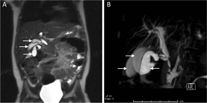
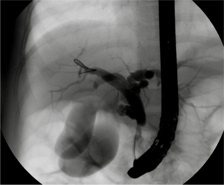
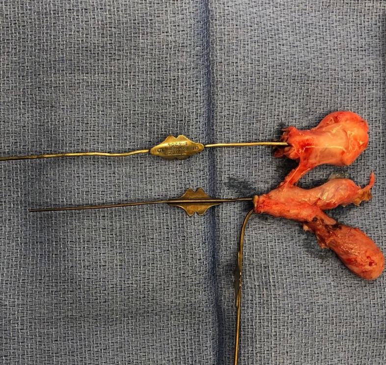
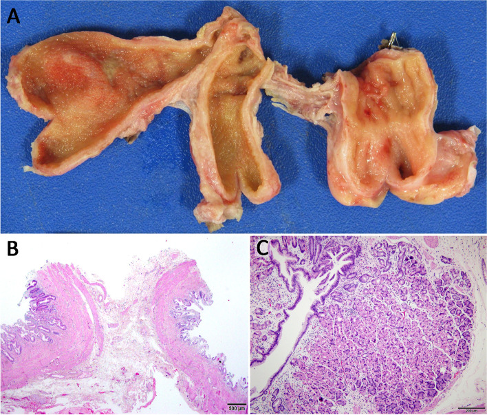
Case presentation: A 2-year-old patient presented to our institution with intermittent abdominal pain for 1 year. She had elevated transaminases with imaging concerning for a choledochal cyst. After assessment with magnetic resonance cholangiopancreatography and endoscopic retrograde cholangiopancreatography, she was diagnosed with a gallbladder multiplication and a common bile duct stricture. She underwent laparoscopic cholecystectomy, which confirmed the diagnosis of triple gallbladder. One of the three gallbladders demonstrated heterotopic gastric mucosa on final pathology, including at the cystic duct margin. Follow up testing with a technetium 99 m scan demonstrated a subtle focus of increased activity in the right upper abdomen at the expected location of the common bile duct, concerning for the presence of residual gastric mucosa. The patient remains well without abdominal pain.

Conclusions: We describe the first case of heterotopic gastric mucosa in a triple gallbladder in a young patient presenting with chronic abdominal pain. We also demonstrate the safety and feasibility of laparoscopic cholecystectomy in young children with triple gallbladder. Finally, we propose an interdisciplinary approach to the management of common bile duct strictures in the setting of ectopic acid secretion, involving a combination of medical management, endoscopic intervention, and possible salvage laparoscopic Roux-en-Y hepaticojejunostomy.

Keywords: Heterotopic gastric mucosa; Multiple gallbladders; Triple gallbladder.


Conflict of interest statement

The authors declare that they have no competing interests.

Figures

Fig. 1
Fig. 1
A Coronal view of preoperative magnetic resonance cholangiopancreatography demonstrating the gallbladder and two additional cystic structures in the gallbladder fossa (white arrows) communicating with biliary tract. B Oblique view of biliary tree reconstruction
Fig. 2
Fig. 2
ERCP fluoroscopy image demonstrating opacification of two separate lumens, consistent with gallbladder duplication
Fig. 3
Fig. 3
Triple gallbladder surgical specimen, with lacrimal probes within each cystic duct
Fig. 4
Fig. 4
A Formalin fixed cholecystectomy specimen with lumens exposed. Conjoined gallbladders 1 and 2 (left and center) showed typical tan velvety biliary type mucosa, whereas attached mass lesion (right) notable for luminal gastric features; pale coarse rugal folds. B Hematoxylin and eosin (H&E) stained microscopic section (20X magnification) demonstrating back-to-back walls of conjoined gallbladders 1 and 2 with typical biliary type mucosa. Mural thickening is present indicating chronic cholecystitis. C Vast majority of separately attached lesion lined by oxyntic (body/fundal) type gastric mucosa (H&E, 100X magnification). Microscopic images obtained using Olympus® BX43 microscope with Olympus® DP27 camera and Olympus® cellSens Entry 2.3 (Build 18,987) image processing software

Similar articles

Cited by

  • Triple gallbladder: radiological review.
    Picchi E, Leomanni P, Dell'Olio VB, Pucci N, Di Giuliano F, Ferrazzoli V, Minosse S, Rho M, Chiocchi M, Garaci F, Floris R. Picchi E, et al. Clin J Gastroenterol. 2023 Oct;16(5):629-640. doi: 10.1007/s12328-023-01829-3. Epub 2023 Aug 11. Clin J Gastroenterol. 2023. PMID: 37563490 Free PMC article. Review.
  • Trifoliate gallbladder: A rare congenital anomaly masquerading as acute cholecystitis.
    Hussain A, Inban P, Akuma CM, Raj R, Akuma O, Nwoke NL, Kelechi AE. Hussain A, et al. Clin Case Rep. 2024 Feb 12;12(2):e8497. doi: 10.1002/ccr3.8497. eCollection 2024 Feb. Clin Case Rep. 2024. PMID: 38348149 Free PMC article.

References

    1. Harlaftis N, Gray SW, Skandalakis JE. Multiple gallbladders. Surg Gynecol Obstet. 1977;145(6):928–934. - PubMed
    1. Roeder WJ, Mersheimer WL, Kazarian KK. Triplication of the gallbladder with cholecystitis, cholelithiasis, and papillary adenocarcinoma. Am J Surg. 1971;121(6):746–748. doi: 10.1016/0002-9610(71)90063-8. - DOI - PubMed
    1. Kato T, Hebiguchi T, Matsuda K, Yoshino H. Action of pancreatic juice on the bile duct: pathogenesis of congenital choledochal cyst. J Pediatr Surg. 1981;16(2):146–151. doi: 10.1016/S0022-3468(81)80341-7. - DOI - PubMed
    1. Schweizer P, Schweizer M. Pancreaticobiliary long common channel syndrome and congenital anomalous dilatation of the choledochal duct--study of 46 patients. Eur J Pediatr Surg. 1993;3(1):15–21. doi: 10.1055/s-2008-1063500. - DOI - PubMed
    1. Singham J, Yoshida EM, Scudamore CH. Choledochal cysts: part 1 of 3: classification and pathogenesis. Can J Surg. 2009;52(5):434–440. - PMC - PubMed

Publication types