. 2022 Jan 20;9(1):4.
doi: 10.1186/s40779-022-00365-4.

Potential roles of vitamin D binding protein in attenuating liver injury in sepsis

Affiliations

Potential roles of vitamin D binding protein in attenuating liver injury in sepsis

Kun Xiao et al. Mil Med Res. .

Abstract

Background: In sepsis, vitamin D binding protein (VDBP) has been shown to be low-expressed. The current study examined the relationship between serum VDBP level and liver injury in sepsis patients, as well as in a mouse model for sepsis and in cultured liver epithelial cell line exposed to lipopolysaccharide (LPS).

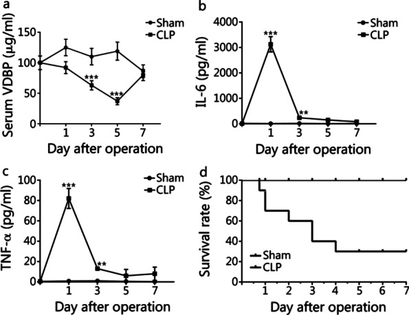
Methods: The human study included 78 sepsis patients and 50 healthy volunteers. Sepsis patients were categorized into sepsis survivor group (n = 43) and sepsis non-survivor group (n = 35) based on 28-day mortality for data analysis. Adult male C57BL/6 mice were subjected to cecal ligation and puncture (CLP). Serum samples were collected on day 1, 3, 5 and 7 to determine the levels of VDBP, 25-hydroxyvitamin D [25(OH)D3], 1,25-dihydroxyvitamin D [1,25(OH)2D3], interleukin-6 (IL-6) and tumor necrosis factor alpha (TNF-α). Potential protective effects of VDBP overexpression against LPS-induced liver damage were examined in cultured THLE2 cells.

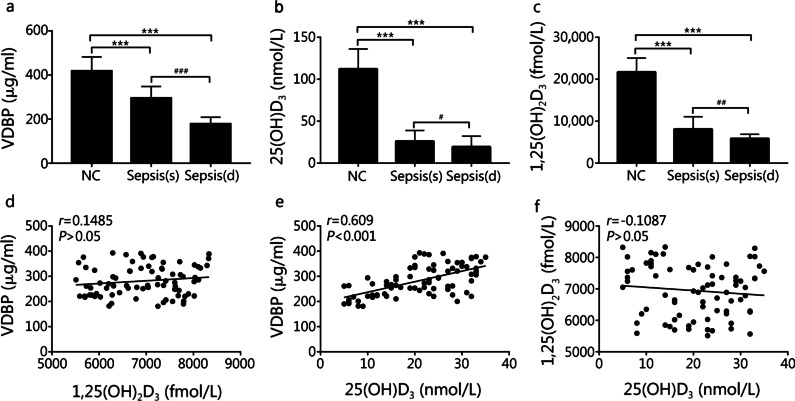
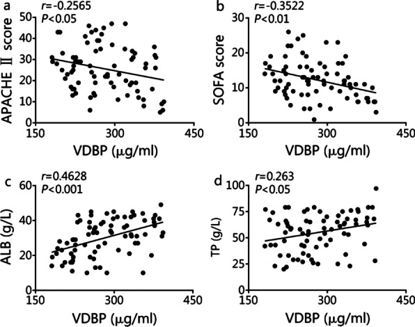
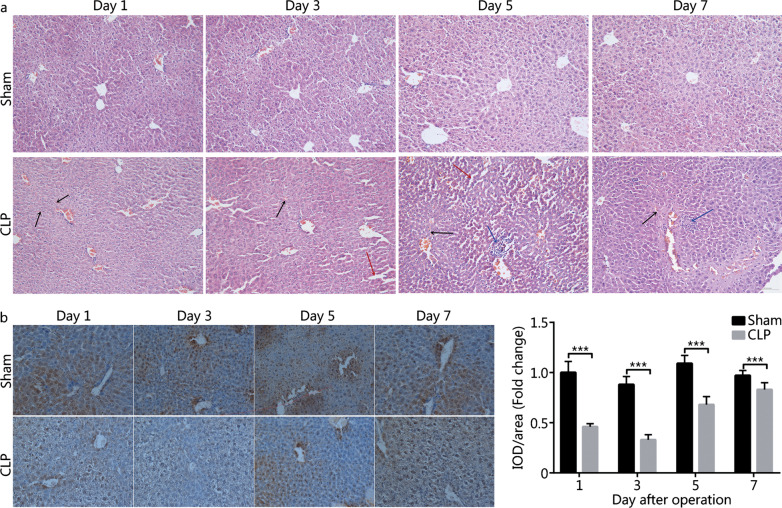
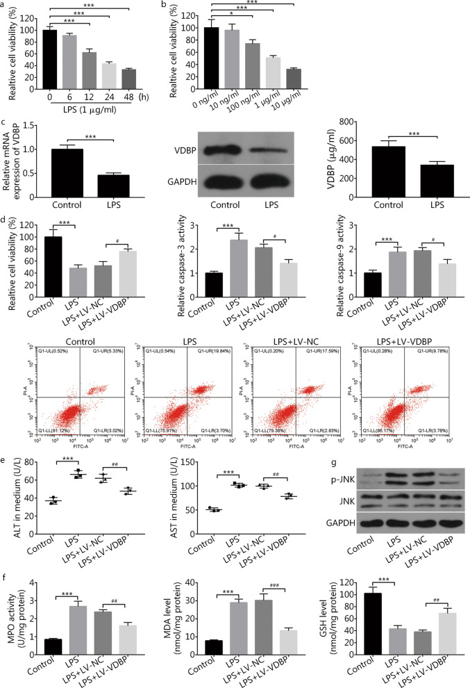
Results: Serum levels of VDBP, 25(OH)D3, and 1,25(OH)2D3 were significantly lower in sepsis patients vs. the healthy control (P < 0.001), as well as in the sepsis non-survivor group vs. the sepsis survivor group (P < 0.001, P = 0.0338, or P = 0.0013, respectively). Lower serum VDBP level was associated with higher Acute Physiology and Chronic Health Evaluation (APACHE) II score (r = - 0.2565, P = 0.0234) and Sequential Organ Failure Assessment score (r = - 0.3522, P = 0.0016), but lower serum albumin (ALB, r = 0.4628, P < 0.001) and total protein (TP, r = 0.263, P = 0.02). In CLP mice, there was a 5-day period of serum VDBP reduction, followed by return towards the baseline on day 7. VDBP was also decreased in LPS-treated THLE2 cells (P < 0.001). VDBP overexpression reduced LPS-induced THLE2 damage. Reduced damage was associated with decreased oxidative stress and inactivation of the c-Jun N-terminal kinase signaling pathway.

Conclusion: VDBP may be protective against sepsis-induced liver injury.

Keywords: Human; Injury; JNK; Liver; Mouse; Sepsis; Vitamin D binding protein.


Conflict of interest statement

The authors have no conflict of interest to declare.

Figures

Fig. 1
Fig. 1
VDBP, 25(OH)D3, and 1,25(OH)2D3 serum levels were notably downregulated in the sepsis patients. ac Serum levels of VDBP, 25(OH)D3, and 1,25(OH)2D3 in healthy volunteer group (NC, n = 50), sepsis non-survivor group [sepsis (d), n = 35], and sepsis survivor group [sepsis (s), n = 43] were measured by using the corresponding ELISA kits. Compared with healthy volunteer group, ***P < 0.001; compared with sepsis survivor group, #P < 0.05, ##P < 0.01 and ###P < 0.001. df Correlation between VDBP and 1,25(OH)2D3 (P > 0.05), VDBP and 25(OH)D3 (P < 0.001), or 1,25(OH)2D3 and 25(OH)D3 (P > 0.05). VDBP vitamin D binding protein
Fig. 2
Fig. 2
Association of VDBP level with disease severity and liver injury. a, b Correlation analysis between VDBP serum level and APACHE II (P < 0.05) or SOFA score (P < 0.01) of sepsis patients (n = 78). c, d Bivariate correlation analysis between VDBP serum level and ALB level (P < 0.001) or TP level (P < 0.05) in sepsis patients (n = 78). ALB albumin, APACHE Acute Physiology and Chronic Health Evaluation, SOFA Sequential Organ Failure Assessment, TP total protein, VDBP vitamin D binding protein
Fig. 3
Fig. 3
Expression analysis of VDBP and inflammatory factors in serum samples of sham mice and CLP mice. Mice (n = 40) were randomly divided into sham group (n = 20) and CLP group (n = 20). Five mice were included at each time point (day 1, 3, 5, 7) after surgery in sham or CLP group. a Mouse VDBP level in serum samples was detected in sham and CLP mice at the indicated time points (day 0, 1, 3, 5, 7) after surgery by mouse VDBP ELISA kit. b, c Mouse IL-6 and TNF-α serum levels were detected in sham and CLP mice at the indicated time points (day 0, 1, 3, 5, 7) after surgery by mouse IL-6 or TNF-α ELISA kit, respectively. d Survival rate of sham and CLP mice. Compared with sham group, **P < 0.01 and ***P < 0.001. CLP cecal ligation and puncture, IL-6 interleukin 6, TNF-α tumor necrosis factor alpha, VDBP vitamin D binding protein
Fig. 4
Fig. 4
Liver pathological analysis and VDBP expression analysis in the liver tissues of CLP mice and sham mice. a HE staining analysis of the liver tissues in sham mice and CLP mice on days 1, 3, 5, and 7 after surgery. Black arrow: hemorrhage; Red arrow: edema and hepatic sinusoid structure abnormality; Blue arrow: inflammatory cell infiltration. b VDBP IHC analysis in the liver tissues of sham mice and CLP mice on days 1, 3, 5, and 7 after surgery. ***P < 0.001 compared with sham group. CLP cecal ligation and puncture, HE hematoxylin–eosin, IHC immunohistochemistry, VDBP vitamin D binding protein, IOD integrated optical density
Fig. 5
Fig. 5
VDBP overexpression weakened LPS-induced THLE2 liver cell injury. a THLE2 cells were treated with 1 μg/ml of LPS. At 0, 6, 12, 24, and 48 h after LPS stimulation, the cell viability was estimated by the MTT assay. Compared with 0 h group, ***P < 0.001. b THLE2 cells were stimulated with different concentrations of LPS for 24 h and then the cell viability was determined by the MTT assay. Compared with 0 ng/ml group, *P < 0.05 and ***P < 0.001. c The THLE2 cells were treated with or without 1 μg/ml of LPS for 24 h. The VDBP mRNA and protein expression levels were measured by RT-qPCR and Western blotting, respectively. The VDBP secretion level was detected by using a commercial kit. Compared with control group, ***P < 0.001. df THLE2 cells infected with recombinant lentiviruses encoding the human VDBP (LV-VDBP) and control (LV-NC) were treated with 1 μg/ml of LPS for 24 h, followed by the detection of cell viability, cell apoptotic rate, caspase-3 activity, caspase-9 activity, ALT, AST, MPO activity, MDA level, and GSH level. Compared with control group, ***P < 0.001; compared with LPS + LV-NC group, #P < 0.05, ##P < 0.01, ###P < 0.001. g THLE2 cells infected with LV-NC or LV-VDBP were treated with 1 μg/ml of LPS for 24 h. The protein levels of p-JNK and JNK were determined by Western blotting. ALT alanine aminotransferase, AST aspartate transaminase, GSH glutathione, JNK c-Jun N-terminal kinase, LPS lipopolysaccharide, MDA malondialdehyde, MPO myeloperoxidase, p-JNK phosphorylated JNK, VDBP vitamin D binding protein

References

    1. Singer M, Deutschman CS, Seymour CW, Shankar-Hari M, Annane D, Bauer M, et al. The third international consensus definitions for sepsis and septic shock (Sepsis-3) JAMA. 2016;315(8):801–810. doi: 10.1001/jama.2016.0287. - DOI - PMC - PubMed
    1. Fleischmann C, Scherag A, Adhikari NK, Hartog CS, Tsaganos T, Schlattmann P, et al. Assessment of global incidence and mortality of hospital-treated sepsis: current estimates and limitations. Am J Respir Crit Care Med. 2016;193(3):259–272. - PubMed
    1. Chousterman BG, Swirski FK, Weber GF. Cytokine storm and sepsis disease pathogenesis. Semin Immunopathol. 2017;39(5):517–528. - PubMed
    1. Van Der Poll T, Van De Veerdonk FL, Scicluna BP, Netea MG. The immunopathology of sepsis and potential therapeutic targets. Nat Rev Immunol. 2017;17(7):407–420. - PubMed
    1. Levi M, Van Der Poll T. Coagulation and sepsis. Thromb Res. 2017;149:38–44. - PubMed

Publication types

Substances