. 2022 Jan;27(1):8-19.
doi: 10.1016/j.slasd.2021.10.012. Epub 2021 Oct 23.

Identification of potent small molecule inhibitors of SARS-CoV-2 entry

Affiliations

Identification of potent small molecule inhibitors of SARS-CoV-2 entry

Sonia Mediouni et al. SLAS Discov. 2022 Jan.

Abstract

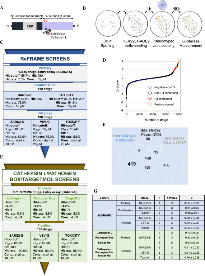
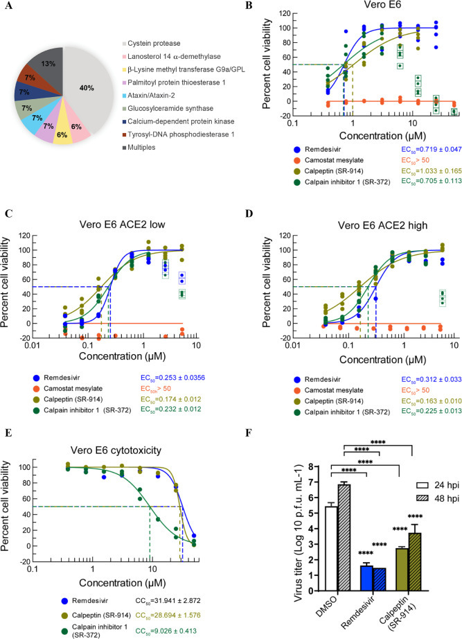
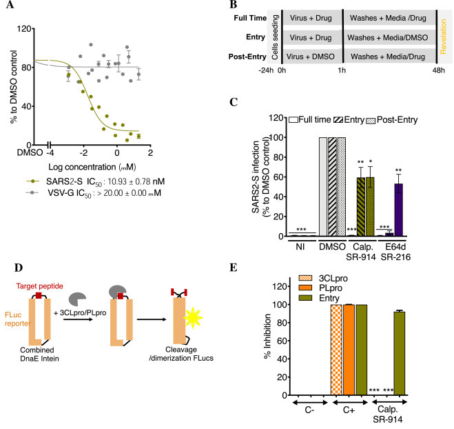
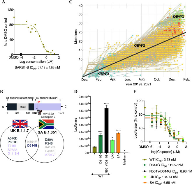
The severe acute respiratory syndrome coronavirus 2 responsible for COVID-19 remains a persistent threat to mankind, especially for the immunocompromised and elderly for which the vaccine may have limited effectiveness. Entry of SARS-CoV-2 requires a high affinity interaction of the viral spike protein with the cellular receptor angiotensin-converting enzyme 2. Novel mutations on the spike protein correlate with the high transmissibility of new variants of SARS-CoV-2, highlighting the need for small molecule inhibitors of virus entry into target cells. We report the identification of such inhibitors through a robust high-throughput screen testing 15,000 small molecules from unique libraries. Several leads were validated in a suite of mechanistic assays, including whole cell SARS-CoV-2 infectivity assays. The main lead compound, calpeptin, was further characterized using SARS-CoV-1 and the novel SARS-CoV-2 variant entry assays, SARS-CoV-2 protease assays and molecular docking. This study reveals calpeptin as a potent and specific inhibitor of SARS-CoV-2 and some variants.

Keywords: Anti-viral drugs; HTS; Inhibitor; SARS-CoV-2; Virus entry.


Conflict of interest statement

Declaration of Conflicting Interest The are no conflicts of interest amongst any of the authors and the work pertained in this manuscript.

Figures

Fig 1
Fig. 1
High-throughput of ReFRAME, Pathogen Box, TargetMol and Cathepsin L drug libraries for SARS-CoV-2 antiviral compounds. A. Schematic of the spike protein of SARS-CoV-2. RBD: receptor binding domain. B. Schematic of the High-throughput assay. Compounds were pre-spotted in 1536-well plates. Next, 2000 HEK293T-ACE2 cells were added to each well and pre-incubated with each compound for 1 h, followed by infection with MLV reporter luciferase virus pseudotyped with the SARS-CoV-2 Spike protein (SARS2-S) or VSV-G protein (VSV-G). Luciferase was measured 48 h later. C. Summary of the ReFRAME library results. Conc.: concentration. D. Distribution of Z-Score for primary screens of each library. Scatter plot of Z_Score for all samples tested from the ReFrame library (N = 1; circle) and other libraries (N = 3; Cathepsin L: square; Pathogen Box: cross; TargetMol: filled circle). Total of 16,320 samples. Positive controls: orange; Negative control: cyan; Hit compounds: red; non-hit compounds: black. E. Summary of the 3 other libraries results. F. ReFrame library screening against different targets: SARS2-S, 3CLpro and PLpro. Venn diagram analysis of comparison between hits from SARS2-S entry, 3CLpro and PLpro assay against ReFRAME library results. There are 419 compounds that are SARS2-entry specific potential inhibitors. G. Robustness in terms of Z’ score of each screen for each library.
Fig 2
Fig. 2
Targets of the selected compounds and SARS-CoV-2 wild type infection. A. Description of the targets of the different hits from all the studied libraries. B. Antiviral activity of the 2 best hits in the SARS-CoV-2-induced CPE assay. Vero E6 cells treated with test compounds for two hours were infected with SARS-CoV-2 at an MOI of 0.05, then incubated for three days in the presence of compound. Cell viability (protection from virus-induced CPE) was measured with CellTiter-Glo. C and D. Antiviral effect was measured with a subset of Vero E6 cells expressing a low (C) and high (D) level of ACE2. E. Cytotoxicity of selected compounds in Vero E6 cells. Cytotoxicity was tested in the same conditions with cell culture media instead of the virus. F. Virus yield reduction activity of selected compounds. Vero E6 cells infected with SARS-CoV-2 at an MOI of 0.05 were cultured in the presence of test compound (5 µM) and the supernatant was harvested after 24 and 48 h of incubation. The Progeny virus was enumerated with a plaque assay using an Avicel overlay in fresh Vero E6 cells. N = 3 experiments were performed for infectivity assays and n = 2 for the cytotoxicity assays. **** P < 0.0001, Two-way ANOVA with Dunnett's multiple comparisons test against DMSO control.
Fig 3
Fig. 3
SR-914 “calpeptin” specifically blocks SARS-CoV entry. A. Its activity against SARS2-S in HEK293T-ACE2-TMPRSS2 cells. Cells were incubated with different concentrations of drugs, then infected with SARS2-S or VSV-G. Luciferase was measured 48 h later, using Bright-Glo. Shown is the mean ± SEM of n = 2 to 4 independent experiments. B. Time of drug addition experiment schematic. Infection was performed for 1 h with or without drugs, Vero CCL81 cells were then washed, and fresh media was added with or without drugs. C. Time of drug addition experiment result. SR-914 was used at 10 µM. E64d at 20 µM. Calp.: calpeptin = SR-914. NI: not infected. Shown is the mean ± SEM of 4 to 6 independent experiments. D. Luciferase complementation assay schematic. The reporter consists of a split Firefly luciferase protein connected by a cleavable peptide for the tested protease. Upon cleavage of the peptide, the luciferase protein undergoes dimerization for an active state. DnaE intein helps in this dimerization. E. Its activity against SARS2-S Entry, 3CLpro and PLpro. C-: negative control. C+; positive control. Shown is the mean ± SD of 3 independent experiments. One-way ANOVA followed by Tukey's post-test were used for statistical comparisons. *, P < 0.01; **, P < 0.001; ***, P < 0.0001.
Fig 4
Fig. 4
Breath of activity of calpeptin against various SARS-CoVs. A. Its activity against SARS1-S in HEK293T-ACE2 cells. HEK293T-ACE2 cells were incubated with different concentrations of calpeptin, then infected with SARS1-S. Luciferase was measured 48 h later, using Bright-Glo. Shown is the mean ± SEM of n = 2 independent experiments. B. Schematic of the substituted residues in the S protein of the highest threat of SARS-CoV-2 strains. C. Evolution of the S protein residues at the position 417, 484, 501 and 614 from 2019 to February 2021. Modified figure from https://nextstrain. org/ncov/global?branchLabel=none&c=gt-S_417,484,501,614&l =clock. D. Activity of the new emergent variants. HEK293T-ACE2 cells were infected with different mutants of SARS2-S. The day after, a medium change was performed. Luciferase was measured 48 h later, using Bright-Glo. Shown is the mean ± SEM of n = 3 independent experiments. WT: wild type, SA: South Africa, UK: United Kingdom. E. Activity of calpeptin activity against crucial mutations present in the S protein of the new emergent strains. Similar experiment than D but calpeptin was added during infection and after medium change. Shown is the mean ± SEM of n = 2–5 independent experiments. Two-way ANOVA followed by Dunnett's post-test were used for statistical comparisons. *, P < 0.01; **, P< 0.001; ***, P < 0.0001.

References

    1. Almaghaslah D., Kandasamy G., Almanasef M., et al. Review on the coronavirus disease (COVID-19) pandemic: its outbreak and current status. Int J Clin Pract. 2020;74:e13637. - PMC - PubMed
    1. Hu B., Guo H., Zhou P., et al. Characteristics of SARS-CoV-2 and COVID-19. Nat Rev Microbiol. 2021;19:141–154. - PMC - PubMed
    1. Cherry J.D. The chronology of the 2002,2003 SARS mini pandemic. Paediatr Respir Rev. 2004;5:262–269. - PMC - PubMed
    1. Mou H., Quinlan B.D., Peng H., et al. Mutations from bat ACE2 orthologs markedly enhance ACE2-Fc neutralization of SARS-CoV-2. BioRxiv. 2020 doi: 10.1101/2020.06.29.178459. https://www.ncbi.nlm.nih.gov/pubmed/32637954 [Online] (accessed Jun 30) - DOI - PMC - PubMed
    1. Moore M.J., Dorfman T., Li W., et al. Retroviruses pseudotyped with the severe acute respiratory syndrome coronavirus spike protein efficiently infect cells expressing angiotensin-converting enzyme 2. J Virol. 2004;78:10628–10635. - PMC - PubMed

MeSH terms

Supplementary concepts

LinkOut - more resources