. 2022 Jan;10(1):e003488.
doi: 10.1136/jitc-2021-003488.

Vectorized Treg-depleting αCTLA-4 elicits antigen cross-presentation and CD8+ T cell immunity to reject 'cold' tumors

Affiliations

Vectorized Treg-depleting αCTLA-4 elicits antigen cross-presentation and CD8+ T cell immunity to reject 'cold' tumors

Monika Semmrich et al. J Immunother Cancer. 2022 Jan.

Abstract

Background: Immune checkpoint blockade (ICB) is a clinically proven concept to treat cancer. Still, a majority of patients with cancer including those with poorly immune infiltrated 'cold' tumors are resistant to currently available ICB therapies. Cytotoxic T lymphocyte-associated antigen-4 (CTLA-4) is one of few clinically validated targets for ICB, but toxicities linked to efficacy in approved αCTLA-4 regimens have restricted their use and precluded full therapeutic dosing. At a mechanistic level, accumulating preclinical and clinical data indicate dual mechanisms for αCTLA-4; ICB and regulatory T cell (Treg) depletion are both thought to contribute efficacy and toxicity in available, systemic, αCTLA-4 regimens. Accordingly, strategies to deliver highly effective, yet safe αCTLA-4 therapies have been lacking. Here we assess and identify spatially restricted exposure to a novel strongly Treg-depleting, checkpoint-blocking, vectorized αCTLA-4, as a highly efficacious and potentially safe strategy to target CTLA-4.

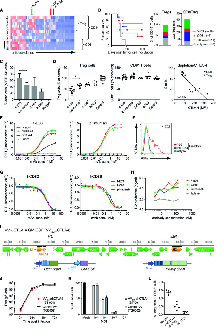
Methods: A novel human IgG1 CTLA-4 antibody (4-E03) was identified using function-first screening for monoclonal antibodies (mAbs) and targets associated with superior Treg-depleting activity. A tumor-selective oncolytic vaccinia vector was then engineered to encode this novel, strongly Treg-depleting, checkpoint-blocking, αCTLA-4 antibody or a matching surrogate antibody, and Granulocyte-macrophage colony-stimulating factor (GM-CSF) (VVGM-αCTLA-4).

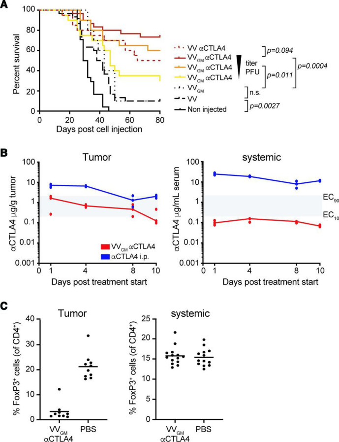
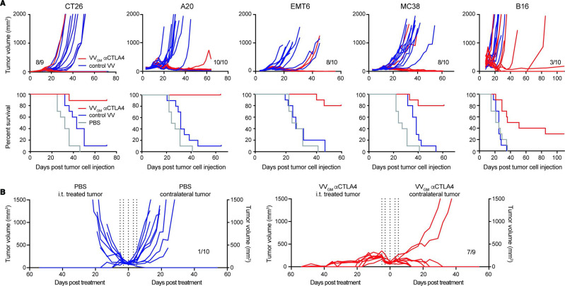
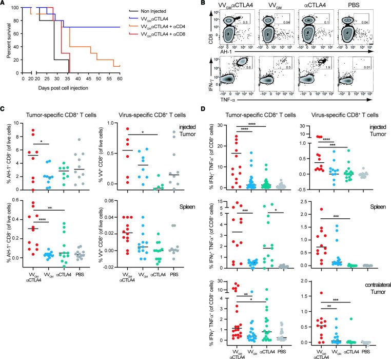
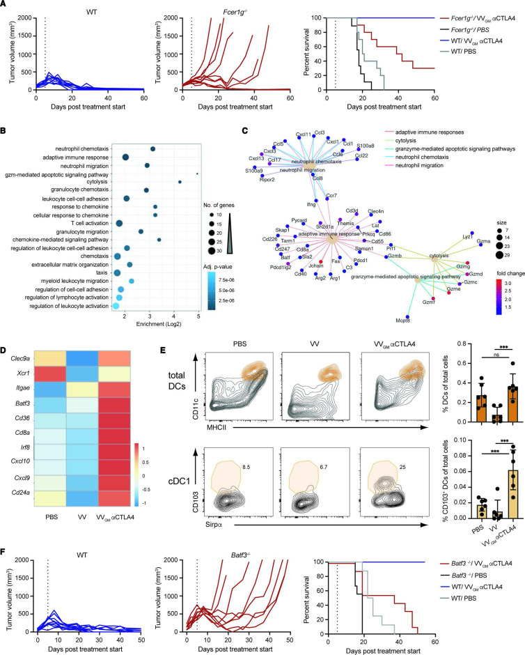
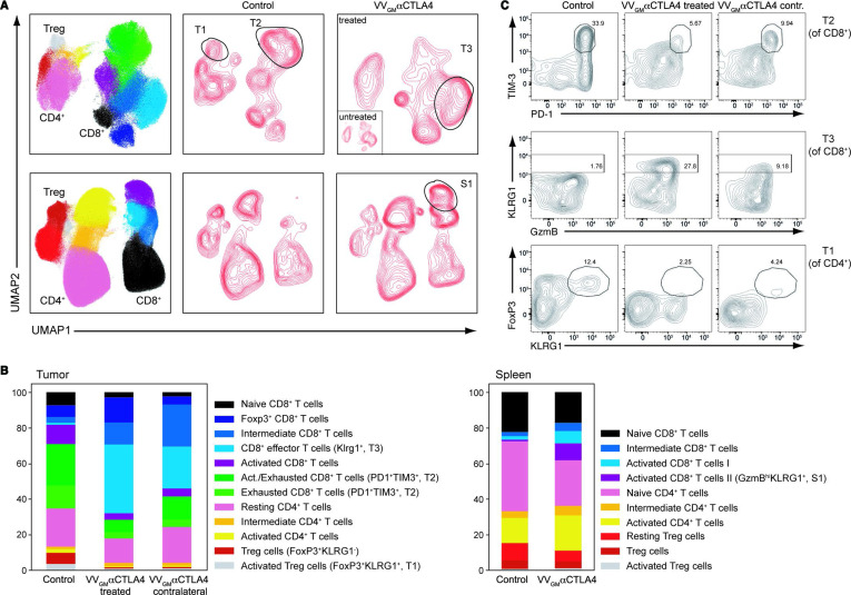
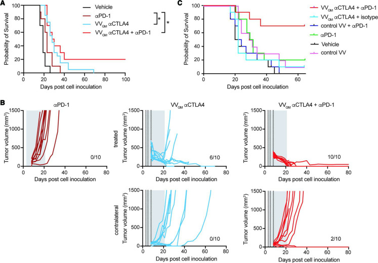
Results: The identified 4-E03 antibody showed significantly stronger Treg depletion, but equipotent checkpoint blockade, compared with clinically validated αCTLA-4 ipilimumab against CTLA-4-expressing Treg cells in a humanized mouse model in vivo. Intratumoral administration of VVGM-αCTLA-4 achieved tumor-restricted CTLA-4 receptor saturation and Treg depletion, which elicited antigen cross-presentation and stronger systemic expansion of tumor-specific CD8+ T cells and antitumor immunity compared with systemic αCTLA-4 antibody therapy. Efficacy correlated with FcγR-mediated intratumoral Treg depletion. Remarkably, in a clinically relevant mouse model resistant to systemic ICB, intratumoral VVGM-αCTLA-4 synergized with αPD-1 to reject cold tumors.

Conclusion: Our findings demonstrate in vivo proof of concept for spatial restriction of Treg depletion-optimized immune checkpoint blocking, vectorized αCTLA-4 as a highly effective and safe strategy to target CTLA-4. A clinical trial evaluating intratumoral VVGM-αhCTLA-4 (BT-001) alone and in combination with αPD-1 in metastatic or advanced solid tumors has commenced.

Keywords: CTLA-4 antigen; antibody specificity; immunotherapy; oncolytic virotherapy.


Conflict of interest statement

Competing interests: MS, MR, PH, LM, CS, FJ, MB, IT, and BF are employees, MS, MR, LM, FJ, MB, IT, and BF are shareholders of BioInvent International. KLC received funding from BioInvent. J-BM, LF, CR, NS, PK, JD, JF, and EQ are employees and shareholders of Transgene.

Figures

Figure 1
Figure 1
Generation and characterization of novel Treg-depleting αCTLA-4 mAbs and oncolytic VVs expressing Treg-depleting αCTLA-4 and GM-CSF. (A) Heatmap shows function-first isolated antibody clones (vertical lines) binding to T cells from CT26 tumor-bearing and naïve BALB/c mice. (B) Antibody-mediated survival (left panel) and TIL modulation (right panel) in CT26 tumor-bearing BALB/c mice. Animals with established tumors received four injections (10 mg/kg) of antibodies with indicated Treg-associated specificity or control mIgG2a antibody (n=5–15). (C) CTLA-4-specific mAbs induce ADCC of in vitro-activated CD4+ T cell. Lysed target T cells were identified by FACS. Figure shows mean±SD (n=4–8); **p<0.01 by Student’s t-test. (D) Anti-CTLA-4 (IgG1) mAbs mediate Treg depletion in vivo in PBMC-humanized mice. Clone 4-E03 shows enhanced depletion of human Treg cells (left panel) but not CD8+ T cells (right panel) compared with ipilimumab. Each dot represents one mouse. Graph shows mean data from two experiments. *p<0.05 by one-way analysis of variance. Right: Level of 4-E03-induced cell depletion plotted in relation to CTLA-4 expression as determined by flow cytometry. (E) 4-E03 hIgG1 and ipilimumab binding to human, mouse, and cynomolgus CTLA-4 and CD28 by ELISA. (F) 4-E03 IgG1 binding to in vitro-activated CTLA-4-expressing human T cells was preblocked with rhCTLA-4-Fc protein (blue line) (G) 4-E03 and 2-C06 block CD80 and CD86 binding to CTLA-4 by ELISA. (H) Functional ligand blockade in vitro. Graphs show interleukin-2 in supernatants following treatment of in vitro activated human PBMCs with αCTLA-4. A representative donor is shown (n=6). (I) Schematic illustration of the VV vectors used to encode heavy (at J2R locus) and light chains of the αCTLA-4 antibody and GM-CSF (at the I4L locus). (J) Replication kinetics in LoVo cells and (K) oncolytic activity on MIA PaCa-2 cells of VVGM-αhCTLA-4 (BT-001). TG6002 (recombinant J2R and I4L deleted VV) was added as control. (L) Functional assessment of αCTLA-4 mAb 4-E03 produced by BT-001-infected MIA PaCa-2 cells in vivo (Treg depletion) as in figure 1D. ADCC, antibody-dependent cell cytotoxicity; FACS, fluorescent-activated cell sorting; ICOS, inducible costimulatory molecule; MOI, multiplicity of infection; PBMC, peripheral blood mononuclear cells; PBS, phosphate-buffered saline; RLU, relative light unit; TIL, tumor-infiltrating lymphocyte; VV, vaccinia virus.
Figure 2
Figure 2
Intratumoral VVGM-αCTLA-4 has in vivo antitumor activity associated with tumor-restricted CTLA-4 receptor saturation and Treg depletion. (A) CT26 tumor-bearing mice were treated with VVGM-αCTLA-4 (7.5×10⁶, 7.5×10⁵, or 7.5×10⁴ pfu), VV-αCTLA-4 (7.5×10⁶ pfu), empty VV (7.5×10⁶ pfu) (n=20–30 mice/group) or VVGM (7.5×10⁶ pfu) (n=10 mice/group). Statistical analysis by log-rank test. (B) Pharmacokinetics of αCTLA-4 in tumors and serum of CT26 tumor-bearing mice after three intratumoral injections (days 0, 2, and 4) of VVGM-αCTLA-4 at 107 pfu or after single intraperitoneal injection of 3 mg/kg of αCTLA-4 mAb 5-B07 (n=3 mice/time point). Area in gray indicates EC10 to EC90 range of CTLA-4 receptor saturation (see online supplemental figure 2A). (C) Numbers of FoxP3+ cells were analyzed by FACS in tumors and spleen at day 10 post VVGM-αCTLA-4 injection. Graphs show pooled data from three independent experiments (n=13 mice/group).
Figure 3
Figure 3
Intratumoral VVGM-αCTLA-4 has broad antitumor activity in syngeneic tumor models spanning inflamed and cold tumor microenvironments. (A) BALB/c mice bearing CT26, A20, or EMT6 tumors, or C57BL/6 mice bearing MC38 or B16 tumors received three intratumoral injections of VVGM-αCTLA-4, control virus lacking αmCTLA-4 mAb (VV empty for A20, EMT6, and MC38 or VVGM for CT26 and B16), or PBS. Treatment started when tumors had a volume of ~50 to 100 mm3. Graphs show tumor growth of individual mice and corresponding survival (n=10). (B) CT26 tumor cells were implanted into the right and left flanks of BALB/c mice. Intratumoral injections (vertical dotted lines, same as in A) in right flank tumors with VVGM-αCTLA-4 started when tumors reached a volume of ~100 mm3 (n=9–10). VV, vaccinia virus.
Figure 4
Figure 4
Intratumoral VVGM-αCTLA-4 elicits robust systemic CD8+ T cell-dependent antitumor immunity. (A) BALB/c mice were treated with CD8 or CD4 depleting antibody pre- and post-subcutaneous challenge with CT26 tumor cells. When tumors reached a volume of ~20 to 50 mm3, treatment as in figure 3A commenced. One representative experiment (out of two) is shown with 10 mice per group. (B–D) CT26 tumor-bearing mice were treated intratumorally with VVs or intraperitoneally with αCTLA-4 mAb (clone 5-B07 at 3 mg/kg). Tumor cell suspensions and splenocytes were restimulated ex vivo with VV-specific or CT26 (AH-1)-specific peptide and the percentage of IFN-γ+ and TNFα+ CD8+ T cells, or MHC class I-labeled multimer positive CD8+ T cells was quantified by FACS. (B) Flow-cytometry dot plots of AH-1 peptide-positive (upper panel) or cytokine-positive (lower panel) splenocytes. Quantification of (C) antigen-specific and (D) IFN-γ+/TNFα+ CD8+ T cells in indicated organs. Each dot represents one mouse (n=3–6 experiments). *p<0.05, **p<0.01, ***p<0.001, ****p<0.0001 by one-way analysis of variance. IFN-γ, interferon gamma; VV, vaccinia virus.
Figure 5
Figure 5
Intratumorally induced CD8+ T cell antitumor immunity is FcγR-dependent and cDC1-dependent. (A) CT26 tumor-bearing WT and Fcer1g−/− BALB/c mice received intratumoral injections of VVGM-αCTLA-4 or PBS as in figure 3A. Graphs show tumor volume (left and center panels) and mouse survival (right panel). Vertical lines indicate the end of the treatment. (n=10 mice/group) (B) GO terms enriched in the set of 352 differentially expressed genes, either upregulated or downregulated, in CT26 tumors treated with VVGM-αCTLA-4 versus VV empty. The 20 enriched terms with the lowest adjusted p value are shown. (C) Network view of the differentially expressed genes associated with the five most enriched GO terms from (B). Only genes upregulated were found associated with these five enriched GO terms. (D) Heatmap representation of cDC1-associated transcripts differentially expressed after treatment with VVGM-αCTLA-4 or VV empty. (E) Representative FACS plots and summarized quantitation of total DCs and cDC1s in tumors following treatment. DCs were gated as described in supplemental information and further defined as CD103+/Sirpα cDC1s. ***p<0.001 by one-way analysis of variance. (F) MC38 tumor-bearing WT and Batf3−/− C57BL/6 mice received intratumoral injections of VVGM-αCTLA-4 or PBS as in figures 3A and 5A. Graphs show tumor volume (left and center panels) and mouse survival (right panel). Vertical lines indicate the end of the treatment. (n=8–10 mice/group).DC, dendritic cell; ns, not significant; VV, vaccinia virus; WT, wild type.
Figure 6
Figure 6
Intratumoral VVGM-αCTLA-4 expands peripheral effector CD8+ T cells and reduces Treg and exhausted CD8+ T cells. CT26 ‘twin’ tumor-bearing BALB/c mice were treated intratumorally (right flank tumors only) with VVGM-αCTLA-4 or PBS. Spleens and injected and contralateral tumors were collected on day 10 post-treatment and stained with a high-dimensional panel designed to identify T-cell populations. (A) Intratumoral VVGM-αCTLA-4 reduced activated CD4+ Treg cells (FoxP3+KLRG1+, ‘T1’), reduced exhausted CD8+ T cells (PD1+TIM3+, and ‘T2’), and expanded activated effector CD8+ T cells (KLRG1+ and ‘T3’) in injected and uninjected tumors (upper panel) and expanded activated CD8+ T cells in spleen (S1, lower panel) (B) shows quantification of data illustrated in A. One representative experiment (out of three) with five mice/group is shown. (C) Flow cytometry plots show characteristic markers of selected intratumoral T-cell clusters. VV, vaccinia virus.
Figure 7
Figure 7
Intratumoral VVGM-αCTLA-4 synergizes with αPD-1 to reject ‘cold’ immune checkpoint blockade-resistant tumors. (A, B) C57BL/6 mice carrying two B16 tumors, one large (5×10⁵ cells, treated tumor) and one small tumor (1×10⁵ cells, contralateral side) received three intratumoral injections with VVGM-αCTLA-4 (vertical dotted lines) and/or intraperitoneal αPD-1 (29F.1A12, 10 mg/kg; two times per week for 3 weeks, gray area). (A) Survival (n=10–20), *p<0.05 by log-rank test. (B) Tumor growth curves of intratumorally injected and contralateral tumors. (C) A20 tumor-bearing BALB/c mice were treated thrice with VVGM-αCTLA-4 intratumorally (at a suboptimal dose of 1×10⁵ pfu), αPD-1 intraperitoneally (RMP1-14, full dose of 10 mg/kg) or the combination of both when tumors had reached a volume of ~135 mm3. Graph shows animal survival (n=10). VV, vaccinia virus.

Similar articles

Cited by

References

    1. Hodi FS, O'Day SJ, McDermott DF, et al. . Improved survival with ipilimumab in patients with metastatic melanoma. N Engl J Med 2010;363:711–23. 10.1056/NEJMoa1003466 - DOI - PMC - PubMed
    1. Larkin J, Chiarion-Sileni V, Gonzalez R, et al. . Combined nivolumab and ipilimumab or monotherapy in untreated melanoma. N Engl J Med 2015;373:23–34. 10.1056/NEJMoa1504030 - DOI - PMC - PubMed
    1. Topalian SL, Hodi FS, Brahmer JR, et al. . Safety, activity, and immune correlates of anti-PD-1 antibody in cancer. N Engl J Med 2012;366:2443–54. 10.1056/NEJMoa1200690 - DOI - PMC - PubMed
    1. Sharma P, Hu-Lieskovan S, Wargo JA, et al. . Primary, adaptive, and acquired resistance to cancer immunotherapy. Cell 2017;168:707–23. 10.1016/j.cell.2017.01.017 - DOI - PMC - PubMed
    1. Chen DS, Mellman I. Oncology meets immunology: the cancer-immunity cycle. Immunity 2013;39:1–10. 10.1016/j.immuni.2013.07.012 - DOI - PubMed

Publication types