. 2022 Jan 20;5(1):74.
doi: 10.1038/s42003-022-03024-5.

Vespakinin-M, a natural peptide from Vespa magnifica, promotes functional recovery in stroke mice

Affiliations

Vespakinin-M, a natural peptide from Vespa magnifica, promotes functional recovery in stroke mice

Hairong Zhao et al. Commun Biol. .

Abstract

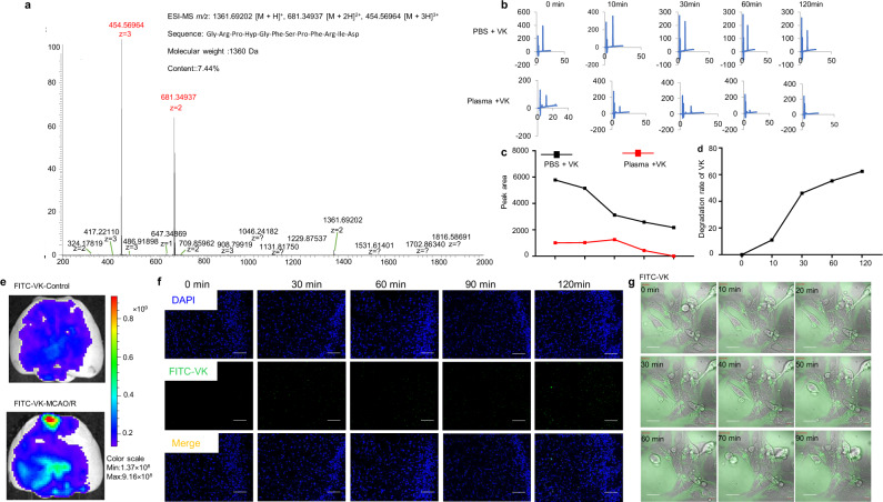
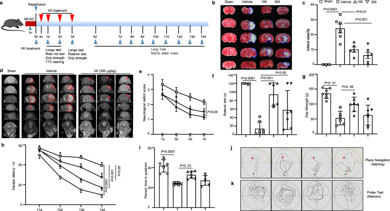
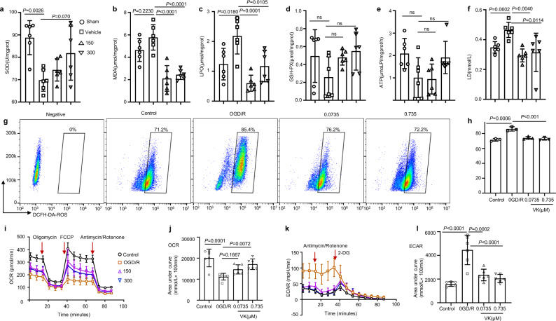
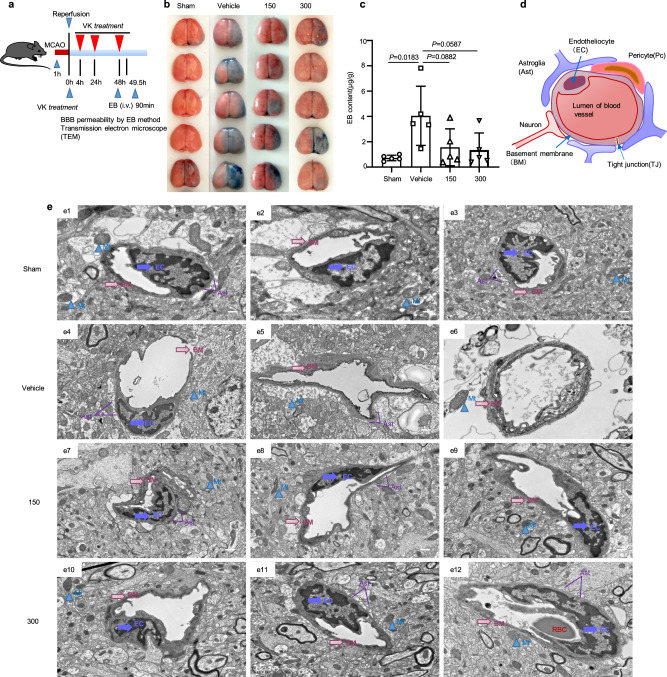
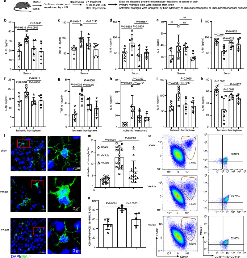
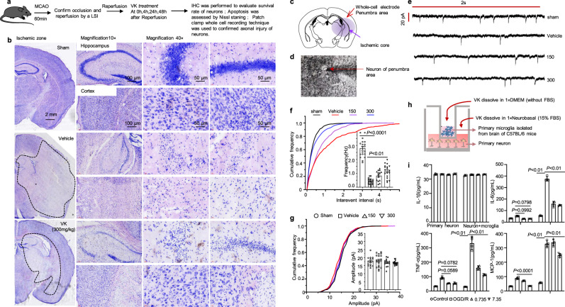
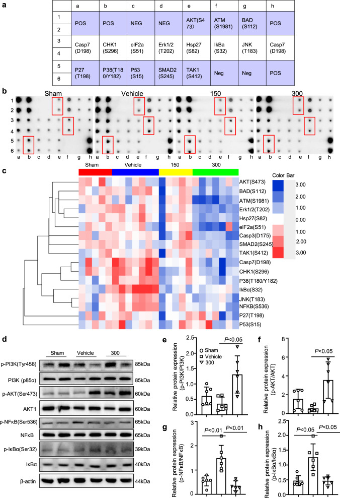
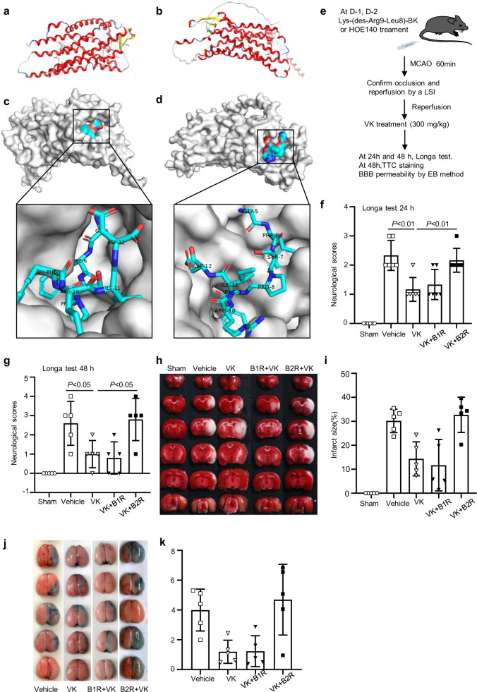
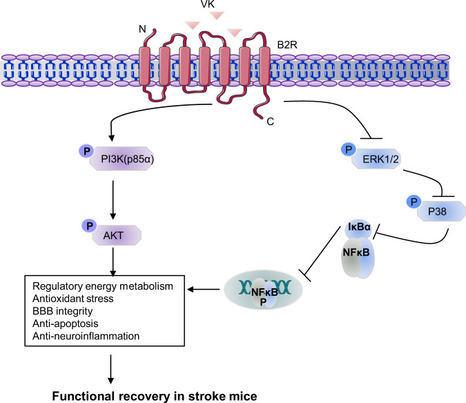
Acute ischemic stroke triggers complex systemic pathological responses for which the exploration of drug resources remains a challenge. Wasp venom extracted from Vespa magnifica (Smith, 1852) is most commonly used to treat rheumatoid arthritis as well as neurological disorders. Vespakinin-M (VK), a natural peptide from wasp venom, has remained largely unexplored for stroke. Herein, we first confirmed the structure, stability, toxicity and distribution of VK as well as its penetration into the blood-brain barrier. VK (150 and 300 µg/kg, i.p.) was administered to improve stroke constructed by middle cerebral artery occlusion in mice. Our results indicate that VK promote functional recovery in mice after ischemia stroke, including an improvement of neurological impairment, reduction of infarct volume, maintenance of blood-brain barrier integrity, and an obstruction of the inflammatory response and oxidative stress. In addition, VK treatment led to reduced neuroinflammation and apoptosis associated with the activation of PI3K-AKT and inhibition of IκBα-NF-κB signaling pathways. Simultaneously, we confirmed that VK can combine with bradykinin receptor 2 (B2R) as detected by molecular docking, the B2R antagonist HOE140 could counteract the neuro-protective effects of VK on stroke in mice. Overall, targeting the VK-B2R interaction can be considered as a practical strategy for stroke therapy.


Conflict of interest statement

The authors declare no competing interests.

Figures

Fig. 1
Fig. 1. Structure, stability, and distribution of VK.
a The matrix-assisted laser desorption/ionization time-of-flight mass spectrometry (MALDI-TOF-MS) profile of Vespakinin-M (VK). The ion at m/z 1361.6854 was the quasi-molecular ion [M + H]+. bd VK (73.5 nM) was incubated for 120 min at physiological pH (7.4) and temperature (37 °C) and the stability of VK in plasma or PBS was continuously detected for 120 min using HPLC. e FITC or FITC-labeled VK were administered intravenously stroke mice and they were killed after 30 min to isolate the brain and imaged by an imaging system (IVIS Lumina II), scale bar = 1 mm. As opposed to red, blue represents the lower permeability of VK. f Brains were collected and cut after VK treatment at 0, 30, 60, 90, and 120 min. Fluorescent images were acquired by confocal microscopy and the distribution of VK was observed, scale bar = 200 µm. g Live-cell imaging was collected in FITC-labeled VK group after OGD/R, scale bar = 20 µm.
Fig. 2
Fig. 2. VK reduced infract volume and promoted sensorimotor and cognitive function in mice subjected to middle cerebral artery occlusion/reperfusion (MCAO/R).
The administration of the VK schedule and behavioral assessment timeline were illustrated schematically (a). Mice (8–10 weeks, males) underwent MCAO for 60 min. The occlusion and reperfusion were confirmed by laser speckle contrast imaging (LSCI). Mice were randomly divided into four groups: sham group, vehicle group, and VK (150 and 300 µg/kg) groups. Mice were administered VK at 0, 4, 22.5, and 46.5 h after MCAO/R as assessed by triphenyl tetrazolium chloride (TTC) staining (b, c) and MRI (d). VK treatment improved sensorimotor recovery as evaluated by the Longa test (e), the rotarod test (f), and the grip test (g) after MCAO/R. The performance in the rotarod test was expressed as the time spent on the rotating rod before falling off and the performance on the grip test was expressed as the score of pullup time for each mouse. Spatial learning and memory were evaluated 11–15 days after MCAO/R by the Morris water maze test. Representative traces indicate the sample paths of the mice from the maze latency trials (learning) (h) and the swimming traces from probe trials (memory) (i). j The latency until the mice located the submerged platform as tested on days 11–14 (defined as spatial learning). k Spatial memory was assessed on day 15 by measuring the time spent swimming in the target quadrant. Statistical analyses were performed by Kruskal–Wallis test with the Dunn post hoc test or two-way repeated-measures ANOVA followed by the Bonferroni post hoc test. Data are expressed as means ± SD, n = 5–7.
Fig. 3
Fig. 3. Effects of VK on oxidative stress and energy metabolism in stroke mice.
a Experimental design: in addition to the sham group, mice (8–10 weeks, male) were subjected to MCAO for 60 min. Mice were randomized into four groups: sham group, vehicle group, and VK (150 and 300 µg/kg) groups. Mice were administered VK at 0, 4, 22.5, and 46.5 h after MCAO/R as assessed by Kits. After reperfusion for 48 h, ischemic tissue was collected. bd The superoxide dismutase (SOD) activity and malondialdehyde (MDA), lipid peroxide (LPO), and glutathione (GSH-PX) levels in the brain were demonstrated by the use of biochemical kits. Adenosine triphosphate (ATP synthase) (e) and lactic acid (LD) (f) were detected to examine the effect of VK treatment on energy metabolism after reperfusion injury. g HT22 cells stained with DCFH-DA for flow cytometry, using without DCFH-DA as a negative control for the FACS gating strategy. h Quantification of DCFH-DA–positive cells as the mean ± SEM from three independent experiments. DCFH-DA (10 μM). i, j Real-time changes in the O2 consumption rate of neurons in response to treatment with the indicated concentrations of VK for 24 h. Cells were treated with 2 μM of oligomycin, 5 μM of carbonyl cyanide-ptrifluoromethoxyphenylhydrazone (FCCP), and 1 μM of rotenone and antimycin, as indicated by the three red arrows. k, l To assess the extracellular acidification rate, cells were treated with 1 μM of rotenone and antimycin and 50 mM of 2-deoxy-d-glucose (2-DG) as indicated by the two red arrows. Statistical analyses were performed using the Kruskal–Wallis test with the Dunn post hoc test or two-way repeated-measures ANOVA followed by Bonferroni’s post hoc test. Data (animal experiment) represent the mean ± SD, n = 5–7. .
Fig. 4
Fig. 4. Protective effect of VK on the blood–brain barrier (BBB) in stroke mice.
a Experimental design: in addition to the sham group, mice (8–10 weeks, male) were subjected to MCAO for 60 min. Mice were randomized into four groups: sham group, vehicle group, and VK (150 and 300 µg/kg) groups. Mice were administered VK at 0, 4, 22.5, and 46.5 h after MCAO/R and BBB integrity was assessed by Evans blue (EB) and transmission electron microscope (TEM). b Brain samples were stained with EB. c EB content in damaged (right) hemisphere was quantized. d Schematic of BBB. The BBB mainly consists of endothelial cells (EC), pericytes (PC), astrocytes (Ast), neurons, tight junctions (TJs), and the basement membranes (BM). The main components of the BBB are cerebral microvascular ECs joined by TJs, thus restricting exogenous molecules into the brain. e The structure of the BBB was observed by TEM. In the sham group (e13), the capillary morphology was regular, the EC and TJ were complete, the thickness of the BM was uniform and continuous around the outside of EC, and the structure of mitochondria (Mt) was clear. When the BBB was destroyed (e46), the BM and TJ were largely dissolved and shed, and the Ast showed extreme edema; there were large electronic blank areas and the Mt was loose or vacuolar, which were relieved by VK treatment (e712). The marks with different symbols indicate the constituent cells or matrix of the BBB. Scale bars: 2 µm. Data are expressed as the mean ± SD, n = 5. Values were analyzed using one-way analysis of variance (ANOVA) with the Tukey multiple comparisons test.
Fig. 5
Fig. 5. VK reduced the release of proinflammatory mediators and inhibited the activation of microglia.
a Experimental design: in addition to the sham group, mice (8–10 weeks, male) were subjected to MCAO for 60 min. Mice were randomized into four groups: sham group, vehicle group, and VK (150 and 300 µg/kg) groups. Mice were administered VK at 0, 4, 24, and 48 h after MCAO/R and the neuroinflammatory response was assessed by ELISA, flow cytometry (FC), immunofluorescence (IF), and immunohistochemical (IHC) analysis. The release of proinflammatory mediators, including IL-1β, TNF, IL-6, and IL-8, in the serum (be) or ischemic cortex (fi) at 48 h after MCAO/R were detected by ELISA. j, k The release of IL-10 in the serum or ischemic cortex was also explored by ELISA at 48 h after MCAO/R. l Microglia were stained with anti-Iba-1 (green; high magnification image from a selected area, green). m Activated microglia were quantified. n Total activated microglia counts. o Total microglia were identified as CD45+F4/80+CD11b+, M1-like microglia were identified as CD45+F4/80+CD11b+ MHCII+. Data are expressed as the mean ± SD, n = 6. Values were analyzed using one-way analysis of variance (ANOVA) with the Tukey multiple comparisons test.
Fig. 6
Fig. 6. VK protects neurons against cell death and axonal injury in stroke mice.
a Experimental design: in addition to the sham group, mice (8–10 weeks, male) were subjected to MCAO for 60 min. Mice were randomized into four groups: sham group, vehicle group, VK (150 and 300 µg/kg) groups. Mice were administered VK at 0, 4, 24, and 48 h after MCAO/R as assessed cell death and axonal injury by IHC staining for NeuN+, Nissl staining, and patch-clamp whole-cell recording. b Nissl staining for the mice hippocampus (CA1 region, interaural: 2.10–2.58 mm, bregma −1.46 to −1.22 mm) and cortex in each group was displayed. Position of stimulating and recording electrodes for conduction velocity measurements in (c, d). e Representative firing patterns of pyramidal neurons from mouse neocortex elicited by depolarizing current steps after MCAO/R. All miniature excitatory post synaptic currents (mEPSCs) were recorded at a holding potential of −65 mV. f Cumulative frequency plots of the interevent interval (left) and quantitative analysis of the frequency of AMPA receptor-mediated mEPSCs (right). g Cumulative frequency plots of the amplitude (left) and quantitative analysis of the amplitude of AMPA receptor-mediated mEPSCs (right). Results are expressed as the mean ± SEM, n = 5–7. Statistical significance was determined by one-way ANOVA and Bonferroni test as post-hoc comparisons. h Schematic showing that primary cortical neurons obtained from fetal C57BL/6 mice of embryonic day 16–17.5 and primary microglia isolated from C57BL/6 mice at postnatal day 1–2 were co-cultured with or without VK. For the co-culture system, (i) cell supernatant was collected to measure proinflammatory factors. Data are mean ± SD (n = 4).
Fig. 7
Fig. 7. Reduced neuroinflammation and apoptosis by VK treatment were associated with PI3K–AKT-mediated NF-κB inhibition.
a Layout of antibody array. b Scanned image of antibody microarray and the protein expression levels were tested with antibody microarray analysis (c). dh Western blot analysis revealed that there were changes in the phosphorylation levels of PI3K (p85α), AKT, IκBα, and NFκB in the VK groups compared to the vehicle group.
Fig. 8
Fig. 8. VK-B1R or B2R interaction and the effect of B1R antagonist or B2R antagonist on stroke in mice.
a Secondary structure of the B1R predicted by AlphaFold (ID: AF-Q61125-F1). b The peptide docking model of VK and B1R with the highest score. c Secondary structure of the B2R predicted by AlphaFold (ID: AF-P32299-F1). α helixes, β sheets, and loops were depicted in red, yellow, and blue, respectively. d The peptide docking model of VK and B2R with the highest score (performed by online HDOCK SERVER: http://hdock.phys.hust.edu.cn/). e Experimental design: Mice (8–10 weeks, male) were randomized into five groups: sham group, vehicle group, VK (300 µg/kg) group, B1R antagonist + VK group, and B2R antagonist + VK group. The B1R + VK and B2R + VK groups were administrated Lys-(des-Arg9-Leu8)-BK and HOE140, respectively, before MCAO-induced stroke. In addition to the sham group, all mice were subjected to MCAO for 60 min. Mice were administered by VK at 0, 4, 24, and 48 h after MCAO/R and the stroke outcomes were assessed by Longa test, TTC staining, and Evans blue (EB) method. f, g The inhibitors affected neurological function following MCAO using the Longa test. h TTC staining of representative coronal sections at 48 h after reperfusion. i Quantitative analysis of infarct size at 48 h after MCAO/R. j Representative coronal brain sections showing EB extravasation. k EB extravasation was detected by fluorescence spectrophotometry. Data are expressed as the mean ± SD, n = 5–6. Data were analyzed using one-way analysis of variance (ANOVA) with the Tukey multiple comparisons test.
Fig. 9
Fig. 9. Neuroprotective effects of VK-B2R interaction in cerebral ischemia.
Targeting the VK–B2R interaction promotes functional recovery in mice after ischemia stroke, including an improvement of neurological impairment, reduction of infarct volume, maintenance of blood-brain barrier integrity, and an obstruction of the inflammatory response and oxidative stress. VK treatment led to reduced neuroinflammation and apoptosis associated with the activation of PI3K–AKT and inhibition of IκBα–NF-κB signaling pathways.

References

    1. Wu S, et al. Stroke in China: advances and challenges in epidemiology, prevention, and management. Lancet Neurol. 2019;18:394–405. - PubMed
    1. Collaborators GBDCoD. Global, regional, and national age-sex specific mortality for 264 causes of death, 1980–2016: a systematic analysis for the global burden of disease study 2016. Lancet. 2017;390:1151–1210. - PMC - PubMed
    1. Lo EH. A new penumbra: transitioning from injury into repair after stroke. Nat. Med. 2008;14:497–500. - PubMed
    1. Dwyer TA, Earl DE, Wang L. The utility of a new in vitro model of the stroke penumbra. J. Neurosci. 2008;28:6537–6538. - PMC - PubMed
    1. Prabhakaran S, Ruff I, Bernstein RA. Acute stroke intervention a systematic review. JAMA. 2015;313:1451–1462. - PubMed

Publication types