. 2022 Jan 20;5(1):73.
doi: 10.1038/s42003-021-02903-7.

Cooperative allostery and structural dynamics of streptavidin at cryogenic- and ambient-temperature

Affiliations

Cooperative allostery and structural dynamics of streptavidin at cryogenic- and ambient-temperature

Esra Ayan et al. Commun Biol. .

Abstract

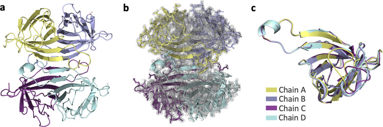
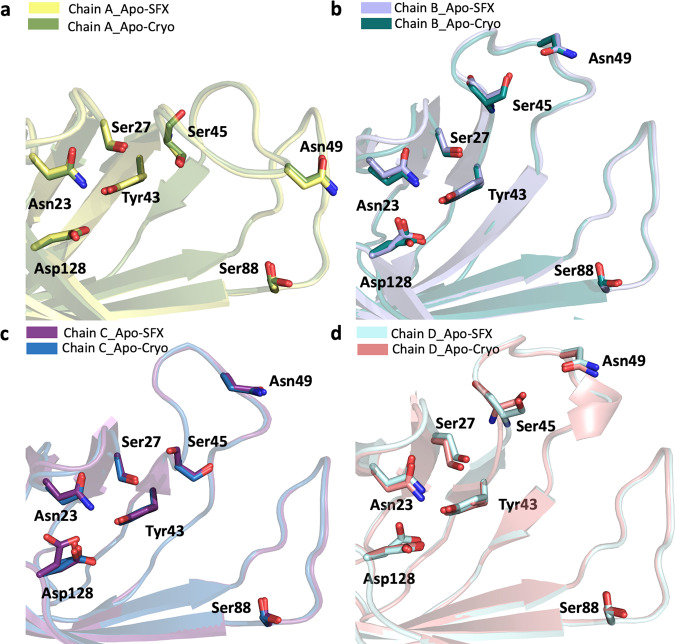
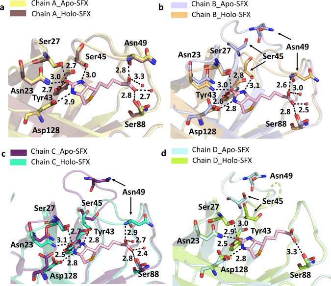
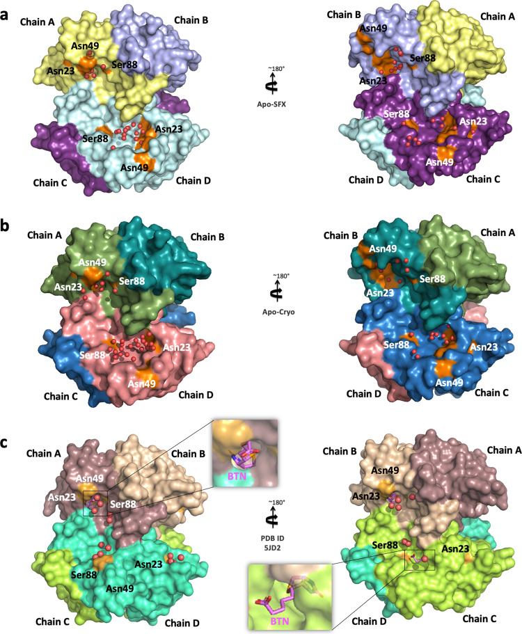
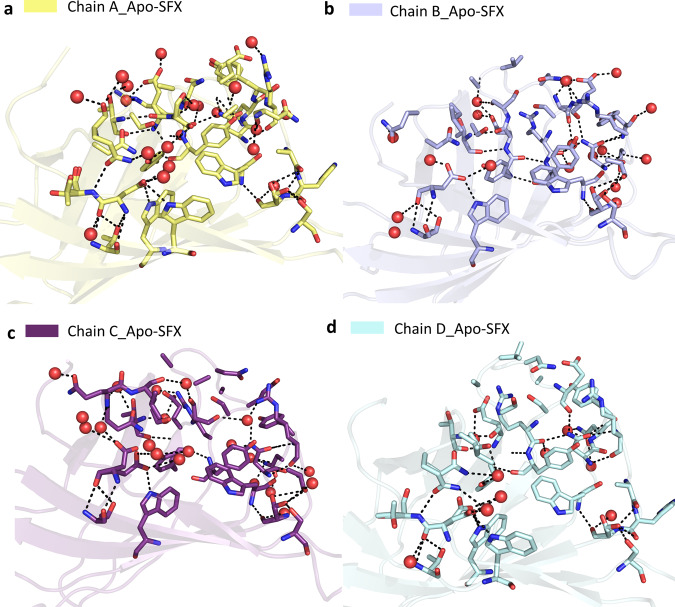
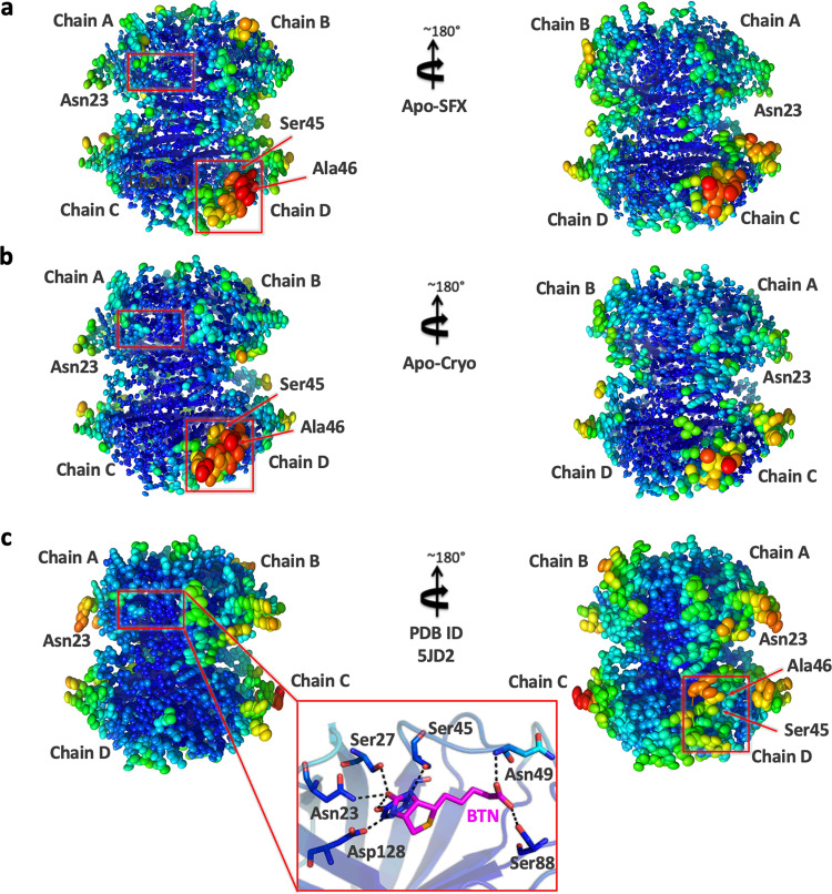
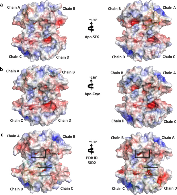
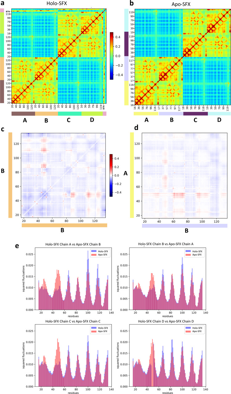
Multimeric protein assemblies are abundant in nature. Streptavidin is an attractive protein that provides a paradigm system to investigate the intra- and intermolecular interactions of multimeric protein complexes. Also, it offers a versatile tool for biotechnological applications. Here, we present two apo-streptavidin structures, the first one is an ambient temperature Serial Femtosecond X-ray crystal (Apo-SFX) structure at 1.7 Å resolution and the second one is a cryogenic crystal structure (Apo-Cryo) at 1.1 Å resolution. These structures are mostly in agreement with previous structural data. Combined with computational analysis, these structures provide invaluable information about structural dynamics of apo streptavidin. Collectively, these data further reveal a novel cooperative allostery of streptavidin which binds to substrate via water molecules that provide a polar interaction network and mimics the substrate biotin which displays one of the strongest affinities found in nature.


Conflict of interest statement

The authors declare no competing interests.

Figures

Fig. 1
Fig. 1. Apo-SFX structure of streptavidin.
a The Apo structure of streptavidin is colored based on each chain. b 2Fo-Fc simulated annealing-omit map at 1 sigma level is colored in gray. c Each chain of streptavidin is superposed with an overall RMSD of 0.177 Å.
Fig. 2
Fig. 2. Biotin-binding site comparison of Apo-SFX and Apo-Cryo structures of streptavidin.
Chains A–D of Apo-SFX is superposed with Apo-Cryo structure in ad, respectively (Supplementary Table 1). There are no significant conformational differences between the two structures.
Fig. 3
Fig. 3. Superposition of the biotin-binding sites for each chain of the Apo-SFX and Holo-SFX structures.
Chains A–D of Apo-SFX is superposed with the Holo-SFX (PDB ID:5JD2) structure of streptavidin in ad, respectively (Supplementary Table 1). The L3/4 opening as a “lid” without selenobiotin binding. Binding of selenobiotin is not symmetric for all four monomers, which represent cooperativity. Selenobiotin and water molecules were represented by light pink sticks and red-colored spheres, respectively. Hydrogen bonds are shown with black dashed lines and their corresponding distance as a unit of Angstrom (Å).
Fig. 4
Fig. 4. Representation of water molecules in the binding pocket of streptavidin structures.
a Apo-SFX structure and b Apo-Cryo structure of apo-state streptavidin and c Holo-SFX structure with selenobiotin (BTN) (PDB ID: 5JD2) are shown with their surface and water molecules are shown with red spheres. The indicated residues around the binding pocket are shown in orange color.
Fig. 5
Fig. 5. Representation of coordinated water molecules and polar interactions near the binding sites for each chain of Apo-SFX structure.
Coordinated water molecules within the binding pocket were altered and polar interactions were reduced with loop opening in Apo-SFX structure. All polar interactions were observed within 3.6 Å. a Binding site residues of chain A were observed with 19 water molecules and 51 polar interactions. Those interactions provide the stability of the chain, which was similar to the selenobiotin-bounded structure. b Chain B-binding site residues were determined with 19 water molecules and 46 polar interactions that were involved. c Residues of the binding site of chain C have 40 polar interactions between residues and 20 water molecules. d Chain D-binding site residues involve 9 water molecules and 31 polar interactions. All interactions included H-bonds and electrostatic interactions and are presented with dashed lines. Water molecules are indicated with red-colored spheres.
Fig. 6
Fig. 6. Representation of thermal ellipsoid structures of streptavidin structures.
a Apo-SFX structure of streptavidin, b Apo-Cryo structure of streptavidin, and (c) Holo-SFX streptavidin in complex with selenobiotin (PDB ID: 5JD2) are shown to determine the stability. Red boxes indicate flexible (red/orange) and stable (blue/green) regions on the streptavidin structures via B-factor presentation. The stable binding pocket of biotin is shown in c.
Fig. 7
Fig. 7. Representation of electrostatic surfaces.
a Apo-SFX structure of streptavidin, b Apo-Cryo structure of streptavidin, and c Holo-SFX streptavidin in complex with selenobiotin (PDB ID: 5JD2) are shown to detect the charge distribution. Holo-SFX structure is colored in light pink and shown with squares.
Fig. 8
Fig. 8. Gaussian Network Model (GNM) analysis results for Apo-SFX and Holo-SFX (PDB ID: 5JD2) structures of streptavidin with selenobiotin.
a Cross-correlation heat-map from overall GNM modes for Holo-SFX structure. Selenobiotin was indicated with “BTN” in the figure. b Apo-SFX structure GNM analysis heat-map results from overall modes. Highly correlated residue motion was represented with red and anti-correlated residue motions with blue color in a, b. c The differences between intrachain cross-correlations of chain B for Holo-SFX structure over Apo-SFX structure results. d Differences in the interchain cross-correlations of Holo-SFX structure over Apo-SFX structure at cross-sections of chains A and B. Differences in correlations are represented with blue color for decrease and red for increase. e Mean squared fluctuations of each chain of Holo-SFX structure and Apo-SFX structure was calculated from the 10 slowest mode from GNM analysis.

References

    1. Hendrickson, W. A. et al. Crystal structure of core streptavidin determined from multiwavelength anomalous diffraction of synchrotron radiation. Proc. Natl Acad. Sci. USA10.1073/pnas.86.7.2190 (1989). - PMC - PubMed
    1. Lim, K. H., Huang, H., Pralle, A. & Park, S. Engineered streptavidin monomer and dimer with improved stability and function. Biochemistry10.1021/bi2010366 (2011). - PubMed
    1. Freitag S, Trong ILE, Klumb L, Stayton PS, Stenkamp RE. Structural studies of the streptavidin binding loop. Protein Sci. 1997 doi: 10.1002/pro.5560060604. - DOI - PMC - PubMed
    1. Bleuler-Martinez S, Schmieder S, Aebi M, Künzler M. Biotin-binding proteins in the defense of mushrooms against predators and parasites. Appl. Environ. Microbiol. 2012 doi: 10.1128/AEM.02286-12. - DOI - PMC - PubMed
    1. Wu SC, Ng KKS, Wong SL. Engineering monomeric streptavidin and its ligands with infinite affinity in binding but reversibility in interaction. Proteins. 2009 doi: 10.1002/prot.22446. - DOI - PubMed

Publication types

MeSH terms