Meta-Analysis
. 2022 Feb;23(2):318-329.
doi: 10.1038/s41590-021-01121-x. Epub 2022 Jan 20.

The immunoregulatory landscape of human tuberculosis granulomas

Affiliations
Meta-Analysis

The immunoregulatory landscape of human tuberculosis granulomas

Erin F McCaffrey et al. Nat Immunol. 2022 Feb.

Erratum in

  • Author Correction: The immunoregulatory landscape of human tuberculosis granulomas.
    McCaffrey EF, Donato M, Keren L, Chen Z, Delmastro A, Fitzpatrick MB, Gupta S, Greenwald NF, Baranski A, Graf W, Kumar R, Bosse M, Fullaway CC, Ramdial PK, Forgó E, Jojic V, Van Valen D, Mehra S, Khader SA, Bendall SC, van de Rijn M, Kalman D, Kaushal D, Hunter RL, Banaei N, Steyn AJC, Khatri P, Angelo M. McCaffrey EF, et al. Nat Immunol. 2022 May;23(5):814. doi: 10.1038/s41590-022-01178-2. Nat Immunol. 2022. PMID: 35277696 Free PMC article. No abstract available.

Abstract

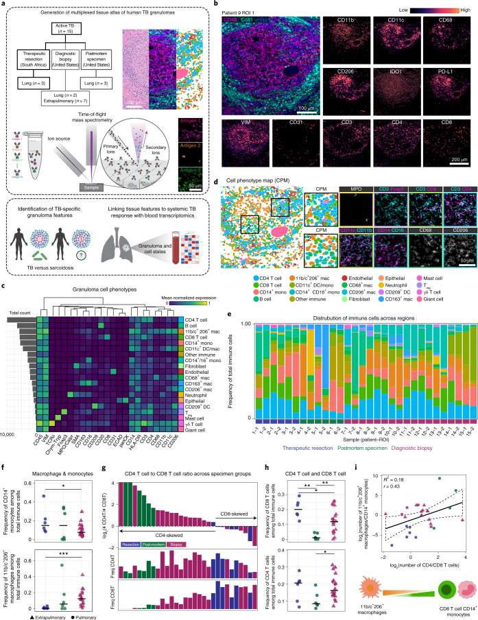
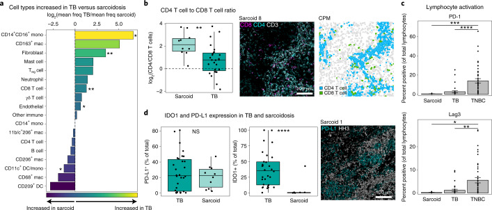
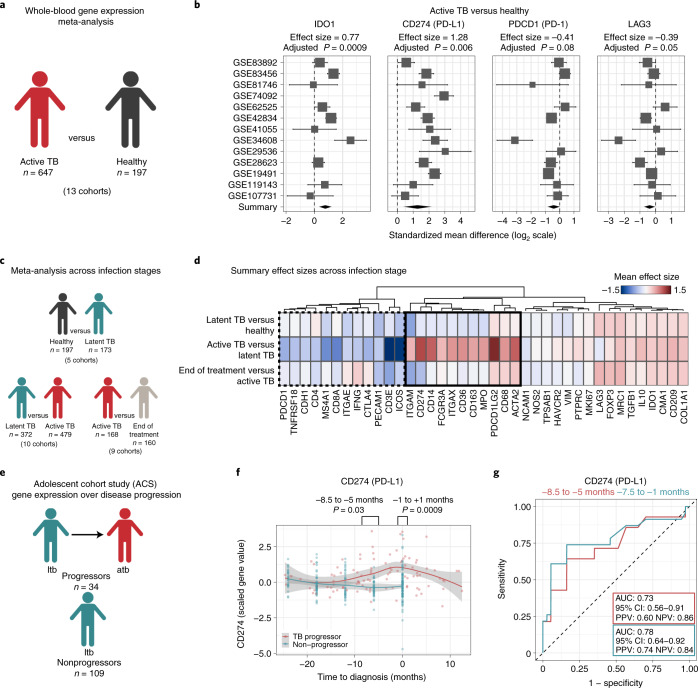
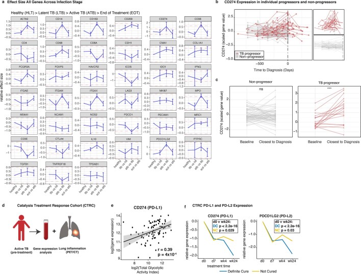
Tuberculosis (TB) in humans is characterized by formation of immune-rich granulomas in infected tissues, the architecture and composition of which are thought to affect disease outcome. However, our understanding of the spatial relationships that control human granulomas is limited. Here, we used multiplexed ion beam imaging by time of flight (MIBI-TOF) to image 37 proteins in tissues from patients with active TB. We constructed a comprehensive atlas that maps 19 cell subsets across 8 spatial microenvironments. This atlas shows an IFN-γ-depleted microenvironment enriched for TGF-β, regulatory T cells and IDO1+ PD-L1+ myeloid cells. In a further transcriptomic meta-analysis of peripheral blood from patients with TB, immunoregulatory trends mirror those identified by granuloma imaging. Notably, PD-L1 expression is associated with progression to active TB and treatment response. These data indicate that in TB granulomas, there are local spatially coordinated immunoregulatory programs with systemic manifestations that define active TB.


Conflict of interest statement

M.A. and S.C.B. are inventors on patent US20150287578A1, which covers the mass spectrometry approach utilized by MIBI-TOF to detect elemental reporters in tissue using secondary ion mass spectrometry. M.A. and S.C.B. are board members and shareholders in IonPath, which develops and manufactures the commercial MIBI-TOF platform. E.F.M. has previously consulted for IonPath. The remaining authors declare no competing interests.

Figures

Fig. 1
Fig. 1. Multiplexed imaging of TB granulomas reveals structured immune cell composition.
a, Conceptual overview of MIBI-TOF analysis of human TB granulomas, comparison with sarcoidosis and complementary analysis of systemic responses to TB. b, Representative images from a TB granuloma. c, Cell lineage assignments based on normalized expression of lineage markers (heatmap columns). Rows are ordered by absolute abundance shown on the bar plot (left), whereas columns are hierarchically clustered (Euclidean distance, average linkage). d, Cell identity overlaid onto the segmentation mask for a representative TB granuloma (left). Two insets (right) are shown. e, The relative abundance of immune cell types across all TB FOVs with cell types ordered by decreasing median abundance and bars ordered by specimen origin (resection, blue; postmortem, green; diagnostic biopsy, red). f, Frequency of CD14+ monocytes and 11b/c+ 206+ macrophages among total immune cells colored by specimen origin. Line represents the median. g, The CD4+ T cell/CD8+ T cell ratio represented as a log2 fold change for each TB FOV (top) colored by specimen origin (top) and frequency of CD4+ T cells (middle) and CD8+ T cells (bottom) among total immune cells. h, Frequency of CD4+ and CD8+ T cells among total immune cells colored by specimen origin. Line represents the median. i, Linear relationship between the CD4+ T cell/CD8+ T cell ratio and 11b/c+ 206+ macrophage/CD14+ monocyte ratio. Linear regression (black solid line) with 95% confidence interval (CI; black dashed line) displayed. Significance was established with a t test (two tailed). Unless specified, all other P values were calculated with a Wilcoxon rank-sum test (two tailed) (*P < 0.05; **P < 0.01; ***P < 0.001). Coll, collagen-1; mac, macrophage; mono, monocyte; MPO, myeloperoxidase; ROI, region of interest; VIM, Vimentin.
Fig. 2
Fig. 2. Spatial analysis of granuloma protein expression and cellular MEs.
a, Positive spatial enrichments (average z-score >0) between protein pairs as a weighted, undirected network (edge weight is proportional to average z-score) with three communities (myeloid core, green; lymphocytic cuff, blue; nonimmune/other, pink). b, Conceptual overview of spatial-LDA. c, Cell probability map (left), max probability map (right), and ME probability for 8 MEs (middle, scaled 0 to 1) for a representative TB granuloma. d, Heatmap of ME preferences for all subsets (standardized mean ME loading) with hierarchical clustering (Euclidean distance, complete linkage) and mean normalized expression of functional markers (probability weighted mean) with columns hierarchically clustered (Euclidean distance, complete linkage). e, Biological classification of MEs. f, Frequency of all MEs per FOV. Heatmap columns are hierarchically clustered (Pearson correlation, complete linkage). Paired ROIs from the same patient annotated with a black bar. ME cluster and sample clinical origin annotated below dendrogram. ExPulm, extrapulmonary; MC, mast cell; pulm, pulmonary; HH3, histone H3; Pan-CK, pan-cytokeratin.
Fig. 3
Fig. 3. Granuloma myeloid cells express a spatially coordinated immunoregulatory program.
a, UMAP visualization of all myeloid populations across all TB FOVs colored by subset (left) and normalized expression of phenotypic markers used to delineate subsets. b, IDO1 and PD-L1 normalized expression overlaid on the UMAP. c, Representative images of TB granulomas showing expression of IDO1 (magenta) and PD-L1 (cyan). d, PD-L1 and IDO1 expression values across all myeloid cells as a biaxial scatter plot. Plot displays Pearson’s r and P value calculated by a t test (two tailed). e, Giant cells identified from a MIBI-scanned TB sample (CD45, green; Vimentin, blue; CD31, red). Representative giant cells displayed in zoomed insets (lower left) with H&E staining or IDO1 (magenta) and PD-L1 (cyan) expression. Bar plot displays the percentage of IDO1+ and PD-L1+ giant cells (n = 34, normalized expression >0). f, The frequency of IDO1+ and PD-L1+ nongranulocytic myeloid cells in aggregate and broken down by ME. Bars represent mean ± s.e.m. (n = 30). g, MEMcore1 and MEIntMono maximum probability maps and representative images of a pulmonary (top) and pleural (bottom) TB sample showing expression of IDO1 (magenta) and PD-L1 (cyan). h, Frequency of PD-L1+ CD163+ macrophages (left) across MEs with a representative MaxPM. Insets are colored by ME (top), cell type (blue, middle) and CD163 (yellow) and PD-L1 (cyan), with the segmentation boundaries overlaid (white). The frequency of IDO1+ CD11c+ DCs (right) across MEs with a representative MaxPM. Insets are colored by ME (top), cell type (green, middle) and IDO1 (magenta), with the segmentation boundaries overlaid (white). Dashed lines represent the total frequency of positive cells (PD-L1 or IDO1) for the indicated cell subset. HLA, human leukocyte antigen; HLA-DR-DQ-DP, HLA-DR/HLA-DQ/HLA-DP.
Fig. 4
Fig. 4. Granuloma lymphocytes display a paradoxical absence of exhaustion markers.
a, Frequency of lymphocyte subsets in all TB FOVs pooled together (left) and representative images of each subset (right). b, Frequency of CD4+ and CD8+ T cells relative to the frequency of total immune cells in four MEs of interest (top). The frequency of Treg cells relative to the frequency of total CD4+ T cells (lower left) (n = 30). c, Frequency of Ki-67+ cells broken down by lymphocyte subset (n = 30). d, Representative image of a TB granuloma, colored by ME assignment (left). Zoomed inset displays Treg cell assignment (upper right: purple, Treg cell; gray, non-Treg cell) and expression of Ki-67 (magenta), CD3 (cyan) and Foxp3 (white) (lower right). e, Percentage of lymphocytes positive for PD-1 (left) and Lag3 (right) in all TB FOVs and TNBC. Bars represent mean ± s.e.m. (TB n = 30, TNBC n = 43). f, The ratio of PD-1+ to PD-L1+ immune cells represented as a log2 fold change in all TB FOVs and TNBC (TB n = 30, TNBC n = 43). Boxplots display the median and interquartile range (IQR; 25–75%) with whiskers representing the upper- and lower-quartile ±1.5× IQR. All P values were calculated with a Wilcoxon rank-sum test (two tailed) (**P < 0.01; ***P < 0.001; ****P < 0.0001).
Fig. 5
Fig. 5. Common and diverging features of immune regulation in TB and sarcoidosis.
a, Fold change of mean frequency of cell subsets (of total cells) in TB versus sarcoidosis with significant differences indicated with an asterisk. b, Comparison of the CD4+ T to CD8+ T cell ratio in TB versus sarcoidosis (TB n = 30, sarcoid n = 10). Representative image of an axillary sarcoidosis FOV showing expression of CD8 (magenta), CD4 (cyan) and CD3 (white) (left) and colored by cell type (right: blue, CD4+ T cell; green, CD8+ T cell). c, Percentage of lymphocytes positive for PD-1 (top) and Lag3 (bottom) in all sarcoidosis FOVs, TB FOVs and TNBC. Bars represent mean ± s.e.m. (sarcoid n = 10, TB n = 30, TNBC n = 43). d, Percentage of total cells positive for IDO1 or PD-L1 in TB and sarcoidosis (TB n = 30, sarcoid n = 10). Representative image of a sarcoidosis FOV showing expression of PD-L1 (cyan) and HH3 (white). Boxplots display the median and IQR (25–75%), with whiskers representing the upper- and lower-quartile ±1.5× IQR. All P values were calculated with a Wilcoxon rank-sum test (two tailed) (NS, P > 0.05; *P < 0.05; **P < 0.01; ***P < 0.001; ****P < 0.0001).
Fig. 6
Fig. 6. Immunoregulatory features of granulomas are reflected in the peripheral blood of TB patients.
a, Conceptual overview of the meta-analysis of patients with active TB (n = 647) versus healthy controls (n = 197). b, Forest plots of gene expression differences in active TB versus healthy individuals. Cohort identifiers are shown on the y axis. Boxes represent the standardized mean difference in gene expression (effect size). The size of the box is proportional to the sample size of that cohort. Whiskers represent the 95% CI, and diamonds (black) represent the overall difference in gene expression between two groups by integrating the standardized mean differences across all cohorts. The width of the diamond corresponds to its 95% CI. The adjusted P values (q values, false discovery rate = 5%) for the summary effect sizes are shown above each plot. c, Conceptual overview of gene expression analysis across clinical infection stage. d, Heatmap of summary gene expression (mean effect size) values in latent TB (n = 173) versus healthy controls (n = 197), latent TB (n = 372) versus active TB (n = 479) and active TB (n = 168) versus end of treatment (n = 160). Clinical stage is displayed on rows, and genes are displayed across columns hierarchically clustered (Euclidean distance, complete linkage). Genes upregulated in active TB versus latent TB are shown in the solid black box, whereas downregulated genes are in the dashed black box. e, Conceptual overview of the ACS. f, PD-L1 gene expression in the ACS cohort across time prior to and after diagnosis of active TB stratified by progressors (red, n = 34) and nonprogressors (blue, n = 109). Gray silhouette represents the 95% CI of the local polynomial regression (red and blue lines). P values were calculated with a Welch two-sample t test. g, Receiver operating characteristic (ROC) curves of the predictive power of CD274 (PD-L1) expression as a predictor of TB progression. atb, active TB; ltb, latent TB; NPV, negative predictive value; PPV, positive predictive value.
Extended Data Fig. 1
Extended Data Fig. 1. Multiplexed imaging of human TB granulomas.
a, Hematoxylin and eosin-stained serial sections of FOVs for MIBI-TOF imaging. b, Multiplexed antibody panel grouped by marker category. c, Grayscale images of endogenous ion signal and proteins in control tissues (tonsil, spleen, placenta). d, Workflow for Deepcell-based segmentation of single cells from multiplexed images. e, Histograms of non-zero signal for all proteins from single-cell data. Blue line represents Gaussian smoothed density fit of histogram. Red line represents automatically identified threshold for marker positivity.
Extended Data Fig. 2
Extended Data Fig. 2. Single-cell phenotypic composition of human TB granulomas.
a, Conceptual overview of hierarchical FlowSOM algorithm application. b, Heatmap of cell lineages clustered by mean normalized protein expression of markers shown along columns. c, Cell phenotype maps for all FOVs. d, Total cell counts across all FOVs sorted by descending order. e, Major cell lineage composition across all FOVs. Bars represent mean ± SEM (n = 30). f, Major cell lineage frequency of total cells broken down by FOV. g, Frequency of major lineages in pulmonary (blue) versus extrapulmonary (grey) TB granulomas. h, Frequency of immune cell subsets (of total immune cells) in pulmonary (blue) versus extrapulmonary (grey) TB granulomas. i, Count of CD14+ monocytes and 11b/c+ 206+ macrophages cells colored by specimen origin. Line represents the median. j, Count of CD4+ and CD8+ T cells colored by specimen origin. Line represents the median. Boxplots display the median and interquartile range (IQR, 25-75%) with whiskers representing the upper- and lower-quartile ± 1.5*IQR. P-values were determined with a Wilcoxon Rank Sum Test (two-tailed) where: * p < 0.05, ** p < 0.01, *** p < 0.001.
Extended Data Fig. 3
Extended Data Fig. 3. Spatial protein enrichment and microenvironment modeling of TB granulomas.
a, Spatial enrichments of protein expression averaged across all TB granuloma FOVs and visualized as a heatmap hierarchically clustered (Euclidean distance, average linkage). Dashed boxes correspond to modules of protein enrichment corresponding to the myeloid core (green), lymphocytic cuff (blue), and a nonimmune/other niche (pink). b, Max probability maps (MaxPM) for all FOVs. c, Representative hematoxylin & eosin and combined myeloid channel of a pleural TB FOV for identification of the myeloid core (left) and frequency of cells in the myeloid core across microenvironments (right). Boxplots display the median and interquartile range (IQR, 25-75%) with whiskers representing the upper- and lower-quartile ± 1.5*IQR (n = 15). d, Counts of cells broken down by phenotype across all FOVs and microenvironments. e, Frequency of cells across microenvironments broken down by specimen type. Line represents the median (n = 30). f, Percent variance explained per clusters based on clustering in Fig. 2f. P-values were determined with a Wilcoxon Rank Sum Test (two-tailed) where: ns p > 0.05, * p < 0.05, ** p < 0.01, *** p < 0.001, **** p < 0.0001.
Extended Data Fig. 4
Extended Data Fig. 4. Immunoregulatory protein expression in TB granulomas.
a, Frequency of cells positive for IDO1 (top) or PD-L1 (bottom) broken down by FOV and cell phenotype. b, Normalized expression of IDO1 (top) and PD-L1 (bottom) for major myeloid subsets ordered by decreasing median expression value. Dashed line indicates the cutoff for positivity for IDO1 (cutoff = 0.26) and PD-L1 (cutoff = 0.25). c, Frequency of IDO1+ or PD-L1+ cells (of total cells) across specimen type (postmortem specimen = green, biopsy = red, therapeutic resection = blue) with dot shape representing organ site (lung = triangle, extrapulmonary = circle). d, Pearson correlation coefficient and p-value determined by t-test (two-tailed) broken down by specimen type. e, Frequency of neutrophils (left) and epithelial cells (right) positive for PD-L1 or IDO1 across all FOVs. f, The count of giant cells across all regions, colored by specimen origin as in c. g, The frequency of IDO1+ and PD-L1+ myeloid cells for all non-granulocytic myeloid cell subsets across ME. Any ME with fewer than 1% of the total cell subset is shaded gray. h, Linear relationship between count of Tregs in Mcore1 with count of IDO1+ (left) and PD-L1+ (right) cells in Mcore1. Linear regression (black solid line) with 95% confidence interval (grey silhouette) displayed. Significance was established with a two-tailed t-test. i, Count of PD-1+ cells across all MEs with >0 positive cells. j The count of IFNγ+ cells in all FOVs with > 0 positive cells, colored by cell type. Unless otherwise specified all p values were determined with a Wilcoxon Rank Sum Test (two-tailed) where: * p < 0.05, ** p < 0.01, **** p < 0.0001.
Extended Data Fig. 5
Extended Data Fig. 5. In situ hybridization of TGFB and IFNG transcripts in human TB granulomas.
a, Representative images from a pulmonary TB granuloma section showing hematoxylin & eosin staining (upper left), MIBI-TOF images (left: CD45 = magenta, VIM = cyan, CD31 = red, αSMA = green, HH3 = blue, right: CD11c = green, PD-L1 = cyan, IDO1 = magenta) and ME assignment with zoomed insets indicated by white or black boxes. b, Representative chromogenic ISH of TB granuloma from a with zoomed inset indicated by black box and colored with fire LUT. c, Quantification of transcripts per area (left) and dots per cell (right). Solid line represents the median for probe (n = 11). Dashed line represents the median DapB signal. d, Quantification of transcripts per cell broken down by granuloma region (orange = myeloid core, pink = lymphocytic cuff). Solid line represents the median for probe (n = 11). Dashed line represents the median DapB signal per region type. e, Representative image of chromogenic ISH in a TB granuloma with zoomed insets indicated by black box. ISH signal colored with fire LUT. f, Quantification of TGFB, IFNG, and DapB transcripts per cell (left) and per area (right). Solid line represents the median for probe (n = 11). g, Proportion IFNγ+ cells measured by MIBI-TOF in MEMcore1 as a fraction of total IFNγ+ cells per ROI. h, Linear relationship between IFNG and TGFB dots per cell (left) and dots per mm2 (right). Linear regression (black solid line) with 95% confidence interval (grey silhouette) displayed. Pearson correlation coefficient displayed. Significance was established with a t-test (two-tailed). Unless specified, all p values were determined with a Wilcoxon Rank Sum Test (two-tailed) where: ns p > 0.05, * p < 0.05, *** p < 0.001, **** p < 0.0001.
Extended Data Fig. 6
Extended Data Fig. 6. In situ hybridization workflow and supplemental analysis.
a, Conceptual overview of combined ISH and MIBI-TOF experimental workflow. b, Representative images of control probes UBC ( + ) and DapB (-) in HeLa cell pellets. c, Representative images of all probes in human spleen and melanoma at 40x magnification. d, Grouped bar plots of total counts (left), area-normalized counts (middle), and cell-normalized counts (right) for all granuloma regions analyzed. e, The ratio of TGFB: IFNG transcripts as a log2 fold-change for total counts (left), area-normalized counts (middle), and cell-normalized counts (right) for all granuloma regions analyzed.
Extended Data Fig. 7
Extended Data Fig. 7. Immunoregulatory protein expression in nontuberculous granulomas.
a, Hematoxylin & eosin-stained sections of sarcoidosis granuloma FOVs. b, Frequency of immune cell subsets out of total immune cells broken down by sarcoidosis FOV. c, Comparison of cell type frequency (out of total cells) between tuberculosis (dark green) and sarcoidosis (light green). Boxplots display the median and interquartile range (IQR, 25-75%) with whiskers representing the upper- and lower-quartile ± 1.5*IQR (TB n = 30, sarcoid n = 10). P-values were determined with a Wilcoxon Rank Sum Test (two-tailed) where: ns p > 0.05, * p < 0.05, and ** p < 0.01. d, Frequency of PD-L1+ cells across all sarcoidosis FOVs broken down by cell subset. e, Representative immunohistochemistry images of PD-L1 or IDO1 (brown) of controls (top = spleen, bottom=placenta), a sarcoid granuloma, xanthoma granuloma, foreign body lesion, and endometrial lesion with hematoxylin nuclear counterstaining (purple). f, Hematoxylin and eosin (left) and MIBI-TOF staining for major cell lineage markers (middle) or IDO1 (magenta) and PD-L1 (cyan) (right) of a representative pulmonary Mycobacterium avium FOV.
Extended Data Fig. 8
Extended Data Fig. 8. Transcriptomic analysis of peripheral blood in TB patients.
a, Gene effect sizes in latent TB (n = 173) versus healthy controls (n = 197), latent TB (n = 372) versus active TB (n = 479), and active TB (n = 168) versus end-of-treatment (n = 160). Bars represent the mean and standard deviation. Dashed red lines represent a relative effect size of 0.6. b, CD274 scaled gene expression in progressors (red) and non-progressors (gray) with zoomed insets displaying groups individually. c, CD274 expression in progressors at the earliest recorded time point prior to progression and the closest time point to diagnosis with ATB (within 30 days). P-values were calculated with a one-sided paired sample t-test. d, Conceptual overview of the Catalysis Treatment Response Cohort (CTRC). e, Correlation between PD-L1 gene expression and total glycolytic activity index (TGAI) represented as log2-transformed values. Linear regression (black line) with 95% confidence interval (grey) displayed. A Pearson correlation of 0.39 (p = 4 ×10⁻⁴) is displayed below the linear fit. Significance was established with a t-test (two-tailed). f PD-L1 (left) and PD-L2 (right) gene expression across treatment time broken down by cure status (blue = definite cure and yellow = no cure). Line represents mean expression in each time point, connected across time points. P-value determined with Student’s t-test for PD-L1 expression at d0 versus wk24 in the definite cure (DC, n = 71) and not-cured (NC, n = 7) groups.

References

    1. World Health Organization. Global Tuberculosis Report 2020https://www.who.int/publications/i/item/9789240013131 (WHO, 2020).
    1. Cohen SB, et al. Alveolar macrophages provide an early Mycobacterium tuberculosis niche and initiate dissemination. Cell Host Microbe. 2018;24:439–446. - PMC - PubMed
    1. Wolf AJ, et al. Mycobacterium tuberculosis infects dendritic cells with high frequency and impairs their function in vivo. J. Immunol. 2007;179:2509–2519. - PubMed
    1. Bold TD, Ernst JD. Who benefits from granulomas, mycobacteria or host? Cell. 2009;136:17–19. - PMC - PubMed
    1. Davis JM, Ramakrishnan L. The role of the granuloma in expansion and dissemination of early tuberculous infectionspan. Cell. 2009;136:37–49. - PMC - PubMed

Publication types

MeSH terms