. 2022 Jan 4:15:744669.
doi: 10.3389/fnbeh.2021.744669. eCollection 2021.

The Perirhinal Cortex Engages in Area and Layer-Specific Encoding of Item Dimensions

Affiliations

The Perirhinal Cortex Engages in Area and Layer-Specific Encoding of Item Dimensions

Nithya Sethumadhavan et al. Front Behav Neurosci. .

Abstract

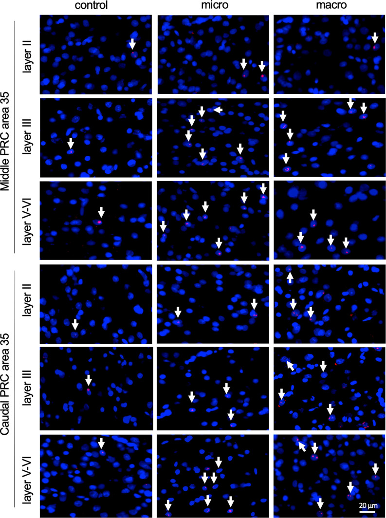
The perirhinal cortex (PRC), subdivided into areas 35 and 36, belongs to the parahippocampal regions that provide polysensory input to the hippocampus. Efferent and afferent connections along its rostro-caudal axis, and of areas 35 and 36, are extremely diverse. Correspondingly functional tasks in which the PRC participates are manifold. The PRC engages, for example, in sensory information processing, object recognition, and attentional processes. It was previously reported that layer II of the caudal area 35 may be critically involved in the encoding of large-scale objects. In the present study we aimed to disambiguate the roles of the different PRC layers, along with areas 35 and 36, and the rostro-caudal compartments of the PRC, in processing information about objects of different dimensions. Here, we compared effects on information encoding triggered by learning about subtle and discretely visible (microscale) object information and overt, highly visible landmark (macroscale) information. To this end, nuclear expression of the immediate early gene Arc was evaluated using fluorescence in situ hybridization. Increased nuclear Arc expression occurred in layers III and V-VI of the middle and caudal parts of area 35 in response to both novel microscale and macroscale object exposure. By contrast, a significant increase in Arc expression occurred in area 36 only in response to microscale objects. These results indicate that area 36 is specifically involved in the encoding of small and less prominently visible items. In contrast, area 35 engages globally (layer III to VI) in the encoding of object information independent of item dimensions.

Keywords: deep layers; fluorescence in situ hybridization; immediate early gene; item encoding; perirhinal cortex; superficial layers; visual information processing.


Conflict of interest statement

The authors declare that the research was conducted in the absence of any commercial or financial relationships that could be construed as a potential conflict of interest.

Figures

Figure 1
Figure 1
Representation of perirhinal cortex areas and micro- and macroscale item paradigms. (A,B) DAPI (4′,6-diamidino-2-phenylindole) stained (blue), nuclei in coronal sections of the rat brain, including outlines highlighting the middle (A) and caudal (B) compartments of the perirhinal cortex (PRC; indicated by white squares). (C,D) Layer organization of the PRC (indicated by white lines). Images were obtained and analyzed in layer II, III, and V-VI/IV-VI of areas 35 and 36 (separated by a yellow line) of the middle (C) and caudal (D) PRC. To simplify the differentiation of the superficial layers, WFS1 (Wolframin, green) was visualized in some sections of the middle and caudal PRC. Nuclei were stained with DAPI (blue). (E,F) Schema of microscale and macroscale item paradigm. Animals participated in a 5 min exploration task, in which small novel objects (microscale items) were placed within three of four holes of a hole board (E), or three large novel objects (macroscale items) were placed on the floor of the chamber (F). (G) Example of manual cell counting to identify somatic Arc mRNA FISH expression in the PRC. Nuclei were identified in an experimenter-blind manner and outlined (white circles). Red signals correspond to Arc mRNA expression. Nuclei that contained an Arc mRNA signal within the DAPI stained area were registered as positively labeled nuclei (indicated by white arrow). Nuclei of glial cells, which are characteristically small and strongly stained with DAPI, were excluded from analysis (top, right).
Figure 2
Figure 2
Exposure to novel microscale or macroscale items significantly enhances nuclear Arc mRNA expression in middle and caudal area 35. (A) Fluorescent image of area 35 showing the superficial layers II and III and the deep layers (separated by white dotted lines), as well as the origin of the representative photomicrographs (white outline). Nuclei were stained with DAPI (blue) and the Arc mRNA signal is shown in red. (B) Photomicrographs showing nuclear Arc mRNA expression (red, indicated by white arrows) in layer III of the caudal area 35 from a control animal (control) and an animal that participated in microscale (micro) or macroscale (macro) item exploration. Blue: nuclear staining with DAPI. Images were taken using a 20x objective. Scale bar: 20 μm. (C) The relative percentage of Arc mRNA positive nuclei in the middle area 35 of control and the two experimental groups (mean ± SEM) is shown. Novel exposure to microscale items (micro, red bar) triggers a significant increase in nuclear Arc mRNA expression in the superficial layer III and deeper cell layers of the middle area 35 compared to their control group (yellow bar). By contrast, novel exposure to macroscale items (macro, blue bar) significantly elevates nuclear Arc mRNA expression in the deep layers of the middle area 35 compared to control animals (Fisher’s LSD post-hoc test: p < 0.05), whereas a tendency is visible for layer III. No significant differences were observed in layer II of the middle area 35. (D) Relative percentage of Arc mRNA positive nuclei in the caudal area 35 of controls and the two experimental groups (mean ± SEM). The exposure to novel objects, regardless of the different dimensions of the novel items, significantly changes nuclear Arc mRNA in the superficial layer III and the deep layers of the caudal area 35 compared to their controls (post-hoc test). In layer II a tendency towards a change can be detected after the exploration of macroscale items (post hoc: caudal control vs. macro p = 0.077845). (C,D) Significant differences for each layer for the experimental groups compared to their control group are marked with asterisks *p < 0.05, **p < 0.01.
Figure 3
Figure 3
Neurons in layer III of area 36 respond to learning about microscale items. (A) Representative DAPI-stained coronal sections of the rat brain showing PRC area 36 and its layer organization (separated by white dotted lines). White squares indicate the origin of the representative photomicrographs. (B) Photomicrographs showing nuclear Arc mRNA expression (red, indicated by white arrows) in layer III of the middle area 36 of control animals (control) or animals that participated in microscale (micro) or macroscale (macro) item exploration. Blue: nuclear staining with DAPI. Images were taken using a 20x objective. Scale bar: 20 μm. (C,D) In the middle area 36 exposure to microscale items (micro) but not macroscale items (macro) triggers a significant increase in Arc mRNA expression in layer III compared to controls (Post hoc test: layer III: middle control vs. micro p < 0.05, caudal control vs. micro p = 0.079281). No changes in Arc mRNA expression were observed in layer II or layers IV-VI of the middle and caudal area 36. The relative percentage of Arc mRNA positive nuclei in the middle (C) and caudal (D) area 36 of the control and the two experimental groups (mean ± SEM). (C,D) Significant differences for each layer for the experimental groups compared to their control group are marked with an asterisk *p < 0.05.
Figure 4
Figure 4
Nuclear Arc mRNA expression in the superficial and the deep layers of area 35 upon exposure to novel items. Photomicrographs showing nuclear Arc mRNA expression (red, indicated by white arrows) in layer II (top), III (middle), and the deep layers (bottom) of the middle and caudal area 35 of control animals (control) or animals that participated in microscale (micro) or macroscale (macro) item exploration. Nuclei (blue) are stained with DAPI. Images were taken using a 20x objective. Scale bar: 20 μm.
Figure 5
Figure 5
Nuclear Arc mRNA expression in the superficial and the deep layers of area 36 following exposure to novel items. Photomicrographs showing Arc mRNA expression (red, indicated by white arrows) in layer II (top row), III (middle row), and the deeper layers (bottom row) of the middle and caudal of area 36 from control animals (control) or animals that participated in microscale (micro) or macroscale (macro) item exploration. Nuclei (blue) are stained using DAPI. Images were taken using a 20x objective. Scale bar: 20 μm.
Figure 6
Figure 6
Hypothesis of projections within hippocampal-perirhinal cortex circuits in novel object learning. Novel acquisition of item information (macroscale, blue) activates neuronal encoding in superficial and deep layers in area 35. Information about novel macroscale items may enter the entorhinal cortex through layers II and III of mostly area 35 before it reaches the hippocampus via the perforant path (Witter et al., ; Doan et al., 2019). In the hippocampus, the dentate gyrus and mossy fibers-CA3 synapses process this information (Kemp and Manahan-Vaughan, ; Hagena and Manahan-Vaughan, ; Hoang et al., 2018). The processed information may then be transferred to the subiculum, which presumably, in turn, sends information back to the deep layers of area 35 (Swanson and Cowan, ; Deacon et al., ; van Groen and Wyss, ; Kloosterman et al., ; Agster and Burwell, 2013). According to our results, area 36 does not support the encoding of this kind of information. If the item dimensions are small (microscale items, red), the Schaffer-collateral-CA1 and commissural-associational (AC)-CA3 synapses process this information (Kemp and Manahan-Vaughan, ; Hagena and Manahan-Vaughan, ; Hoang et al., 2018). Acquisition of microscale items (red) enhances neuronal activity in the superficial layer III of areas 35 and 36. This information may then be sent to the entorhinal cortex which in turn forwards the information to the Schaffer-collaterals-CA1 and AC-CA3 synapses in the hippocampus. In addition, information directly originating from the PRC superficial layers may be of particular importance for microscale information processing in CA1/subiculum (Agster and Burwell, 2013). Finally, the subiculum receives microscale item information that may be sent back to the deep layers of area 35 (Swanson and Cowan, ; Deacon et al., ; van Groen and Wyss, ; Kloosterman et al., ; Agster and Burwell, 2013).

References

    1. Aggleton J. P., Albasser M. M., Aggleton D. J., Poirier G. L., Pearce J. M. (2010). Lesions of the rat perirhinal cortex spare the acquisition of a complex configural visual discrimination yet impair object recognition. Behav. Neurosci. 124, 55–68. 10.1037/a0018320 - DOI - PMC - PubMed
    1. Aggleton J. P., Brown M. W. (2005). Contrasting hippocampal and perirhinal cortex function using immediate early gene imaging. Q. J. Exp. Psychol. B. 58, 218–233. 10.1080/02724990444000131 - DOI - PubMed
    1. Aggleton J. P., Brown M. W., Albasser M. M. (2012). Contrasting brain activity patterns for item recognition memory and associative recognition memory: insights from immediate-early gene functional imaging. Neuropsychologia 50, 3141–3155. 10.1016/j.neuropsychologia.2012.05.018 - DOI - PubMed
    1. Agster K. L., Burwell R. D. (2009). Cortical efferents of the perirhinal, postrhinal and entorhinal cortices of the rat. Hippocampus 19, 1159–1186. 10.1002/hipo.20578 - DOI - PMC - PubMed
    1. Agster K. L., Burwell R. D. (2013). Hippocampal and subicular efferents and afferents of the perirhinal, postrhinal and entorhinal cortices of the rat. Behav. Brain Res. 254, 50–64. 10.1016/j.bbr.2013.07.005 - DOI - PMC - PubMed

LinkOut - more resources