Review
. 2021 Nov 27;13(6):1127-1138.
doi: 10.1007/s12551-021-00858-x. eCollection 2021 Dec.

Protein conformational dynamics and phenotypic switching

Affiliations
Review

Protein conformational dynamics and phenotypic switching

Prakash Kulkarni et al. Biophys Rev. .

Abstract

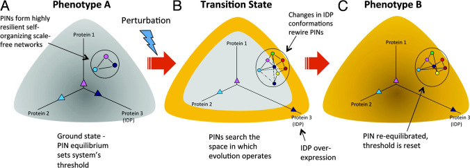
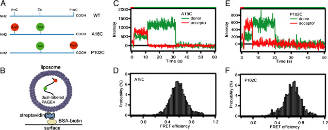
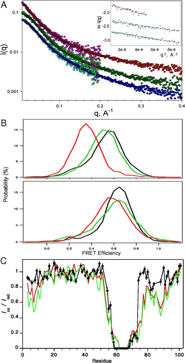
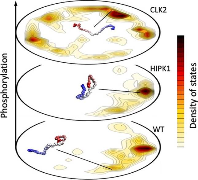
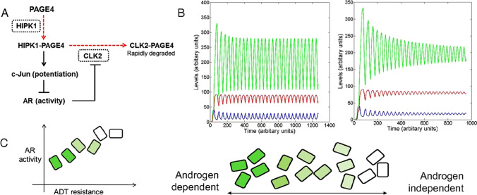
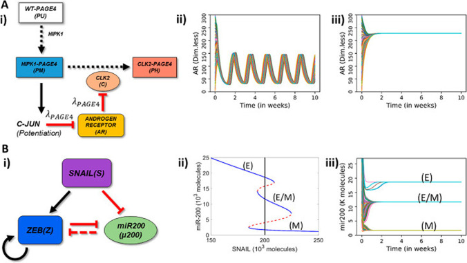
Intrinsically disordered proteins (IDPs) are proteins that lack rigid 3D structure but exist as conformational ensembles. Because of their structural plasticity, they can interact with multiple partners. The protein interactions between IDPs and their partners form scale-free protein interaction networks (PINs) that facilitate information flow in the cell. Because of their plasticity, IDPs typically occupy hub positions in cellular PINs. Furthermore, their conformational dynamics and propensity for post-translational modifications contribute to "conformational" noise which is distinct from the well-recognized transcriptional noise. Therefore, upregulation of IDPs in response to a specific input, such as stress, contributes to increased noise and, hence, an increase in stochastic, "promiscuous" interactions. These interactions lead to activation of latent pathways or can induce "rewiring" of the PIN to yield an optimal output underscoring the critical role of IDPs in regulating information flow. We have used PAGE4, a highly intrinsically disordered stress-response protein as a paradigm. Employing a variety of experimental and computational techniques, we have elucidated the role of PAGE4 in phenotypic switching of prostate cancer cells at a systems level. These cumulative studies over the past decade provide a conceptual framework to better understand how IDP conformational dynamics and conformational noise might facilitate cellular decision-making.

Keywords: Conformational noise; Intrinsically disordered proteins; MRK hypothesis; PAGE4; Phenotypic switching; Protein conformational dynamics.


Conflict of interest statement

Conflict of interest/Competing interestsThe authors declare no competing interests.

Figures

Fig. 1
Fig. 1
Rewiring of protein networks facilitates state-switching by activating latent pathways. (A) The state of a cell with phenotype A is depicted in grey and shows a simple protein network with three proteins (1‒3), of which one is an IDP (indicated in dark blue), and expressed at different levels represented by the three vectors. This configuration represents the protein network’s ground state threshold. (B) Depicts a transition state. A perturbation causes increased IDP expression (protein 3). Overexpression of the IDP results in promiscuity and the protein network explores the network search space shown by the various dashed lines. This transition state is depicted state in yellow around the grey area. (C) The state of the cell after it has transitioned to phenotype B from phenotype A represented in yellow. A particular configuration of the protein network that increased its fitness is “selected,” which now represents the new ground state. Reproduced with permission from Mahmoudabadi et al.
Fig. 2
Fig. 2
Single molecule FRET indicates that PAGE4 is an intrinsically disordered protein. (A) Schematic of the PAGE4 constructs with the native cysteine (green) and the introduced cysteine (red). Single PAGE4 protein molecules were encapsulated inside 100 nm diameter liposomes tethered to a quartz surface. (B) Shows a cartoon of this immobilization scheme (not to scale). Fluorescence emission time courses in the donor and acceptor spectral bands were collected and those indicating exactly 1 donor and 1 acceptor were further analyzed. Example intensity time courses showing anti-correlated donor/acceptor behavior upon photobleaching, which is characteristic of single molecules, are shown for the A18C/63C (C) and P102C/63C (E) FRET mutants. The color bar at the top indicates the illumination color. Red illumination at the start driving only acceptor fluorescence allows identification of molecules containing an active acceptor. The disappearance of red emission (with anticorrelated recovery of green) is photobleaching of the acceptor, and disappearance of green emission is photobleaching of the donor. Histograms assembled from all FRET active data points of over 300 molecules are shown for A18C/63C (D) and P102C/63C (F) PAGE4 mutants. These FRET signals agree with expectations based upon modeling PAGE4 as a highly flexible IDP. Reproduced with permission from Rajagopalan et al.
Fig. 3
Fig. 3
Conformational expansion of PAGE4 upon hyperphosphorylation by CLK2. (A) Experimental X-ray scattering data for the WT-PAGE4 (bottom curve, cyan/blue), HIPK1-PAGE4 (middle curve, light green/dark green), and CLK2-PAGE4 (top curve, pink/red). For each of the variants, the two colors denote independent data collections probing lower-q and medium-q ranges of the scattering data. The curves are offset for clarity. (Inset) Guinier fits of the lowest q data that yield model-free estimates of the ensemble-averaged radii of gyration for the three variants. (B) smFRET measurements. (Upper) Distributions of smFRET efficiency measurements for PAGE4 with donor and acceptor sites at positions 18 and 63 WT-PAGE4 (black), HIPK1-PAGE4 (green), and CLK2-PAGE4 (red). (Lower) Donor and acceptor sites are at positions 63 and 102 for WT-PAGE4 (black), HIPK1-PAGE4 (green), and CLK2-PAGE4 (red). (C) PRE data for CLK2-PAGE4 (black) with an MTSL spin label at C63. Results are compared with earlier observations for WT-PAGE4 (red) and HIPK1-PAGE4 (green). Reproduced with permission from Kulkarni et al.
Fig. 4
Fig. 4
Employing the energy landscape visualization method (ELViM), different PAGE4 ensembles are represented in one single conformational phase space. The density of states, shown in the contour plots, varies according to the physical–chemical conditions, which in this case is the PAGE4 phosphorylation state. Each free energy valley can be characterized by specific conformations that entail particular binding affinities, typical of the promiscuous behavior of IDPs. For WT-PAGE4, through a fly-casting mechanism, the C-terminal region is extended, allowing the binding to its cognate partner. For the HIPK1-PAGE4, the lower free energy of the compact state decreases the affinity for c-Jun. Finally, the dominant extended conformations of CLK2-PAGE4 inhibit any binding affinity
Fig. 5
Fig. 5
Modeling the PAGE4/AP-1/AR/CLK2 regulatory circuit. (A) Regulatory circuit for PAGE4/AP-1/AR/CLK2 interactions. Dashed red lines denote enzymatic reactions, and solid black lines denote non-enzymatic reactions. CLK2 and HIPK1, the two enzymes involved, are shown in dotted rectangles. (B) Dynamics of the circuit showing sustained and damped oscillations for HIPK1-PAGE4 (PAGE4M, shown in blue), CLK2-PAGE4 (PAGE4H, shown in red), and CLK2 (shown in green). (C) Distribution of androgen dependence for an isogenic population over a spectrum, as indicated by the shade of green. Dark green boxes denote highly androgen-dependent (i.e., ADT-sensitive) cells, and white boxes denote androgen-independent cells. Reproduced with permission from Kulkarni et al.
Fig. 6
Fig. 6
Schematic representation of PAGE4-AR and EMT circuits and their stand-alone dynamics. (A) (i) Schematic representation of PAGE4-Androgen Receptor (AR) circuit: The enzyme HIPK1 double phosphorylates WT-PAGE4 and forms the HIPK1-PAGE4 complex which can be further hyperphosphorylated by CLK2 enzyme. Solid arrows show activation, dotted arrows show phosphorylation and red hammer heads show inhibition. In turn, the HIPK1-PAGE4 complex regulates CLK2 levels via the intermediates c-JUN and AR. A strong inhibition of AR by c-JUN and that of CLK2 by AR leads to oscillations (λPAGE4 = 0.1) (ii) or a single steady state (mono-stability) (λPAGE4 = 0.9) (iii). (B) (i) EMT circuit: ZEB and microRNA-200 form a mutually inhibiting loop while SNAIL acts as an external EMT inducer. Solid arrows show transcriptional activation, dashed line show microRNA-mediated inhibition, and solid hammerheads show transcriptional inhibition. (ii) Bifurcation diagram of microRNA (miR)-200 as a function of SNAIL shows tristability, bistability or mono-stability depending on SNAIL levels. Blue and red curves show stable and unstable states respectively. The vertical black line depicts the SNAIL level (= 200,000 molecules) used in panel (iii). (iii) Dynamics of miR-200 for SNAIL = 200 K showing the existence of three states-epithelial (high miR-200; 20 K molecules), mesenchymal (low miR-200; ~ 12 K molecules). In panels A—ii, A—iii, B—iii, different curves depict AR and miR-200 dynamics starting from multiple randomized initial conditions. Reproduced with permission from Singh et al. Entropy (Basel). 2021 Feb 26;23(3):288

References

    1. Al Emran A, Marzese DM, Menon DR, Stark MS, Torrano J, Hammerlindl H, Zhang G, Brafford P, Salomon MP, Nelson N, Hammerlindl S, Gupta D, Mills GB, Lu Y, Sturm RA, Flaherty K, Hoon DSB, Gabrielli B, Herlyn M, Schaider H. Distinct histone modifications denote early stress-induced drug tolerance in cancer. Oncotarget. 2017;9(9):8206–8222. doi: 10.18632/oncotarget.23654. - DOI - PMC - PubMed
    1. Arai M, Sugase K, Dyson HJ, Wright PE. Conformational propensities of intrinsically disordered proteins influence the mechanism of binding and folding. Proc Natl Acad Sci U S A. 2015;112(31):9614–9619. doi: 10.1073/pnas.1512799112. - DOI - PMC - PubMed
    1. Babu MM, van der Lee R, de Groot NS, Gsponer J. Intrinsically disordered proteins: regulation and disease. Curr Opin Struct Biol. 2011;21(3):432–440. doi: 10.1016/j.sbi.2011.03.011. - DOI - PubMed
    1. Babu MM. The contribution of intrinsically disordered regions to protein function, cellular complexity, and human disease. Biochem Soc Trans. 2016;44(5):1185–1200. doi: 10.1042/BST20160172. - DOI - PMC - PubMed
    1. Baggio F, Bozzato A, Benna C, Leonardi E, Romoli O, Cognolato M, Tosatto SC, Costa R, Sandrelli F. 2mit, an intronic gene of Drosophila melanogaster timeless2, is involved in behavioral plasticity. PLoS One. 2013;8(9):e76351. doi: 10.1371/journal.pone.0076351. - DOI - PMC - PubMed

LinkOut - more resources