Review
. 2021 Dec 31:86:e692-e699.
doi: 10.5114/pjr.2021.112377. eCollection 2021.

COVID-19 pandemic in flu season. Chest computed tomography - what we know so far

Affiliations
Review

COVID-19 pandemic in flu season. Chest computed tomography - what we know so far

Marcin Stański et al. Pol J Radiol. .

Abstract

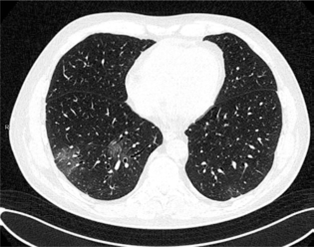
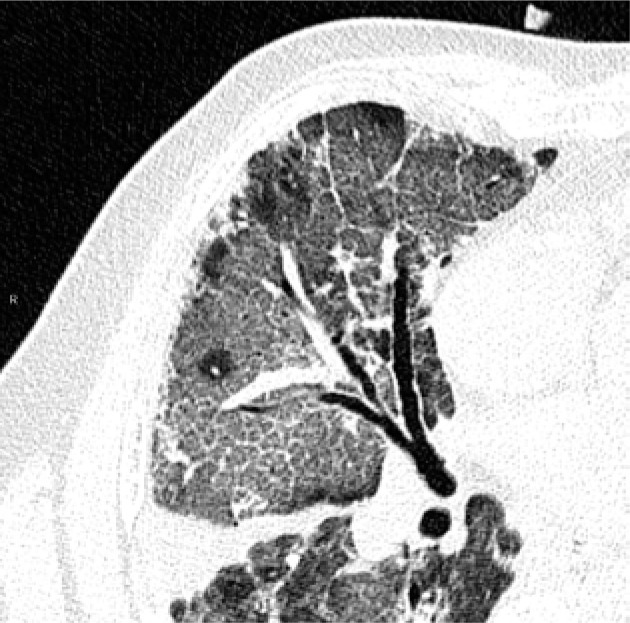
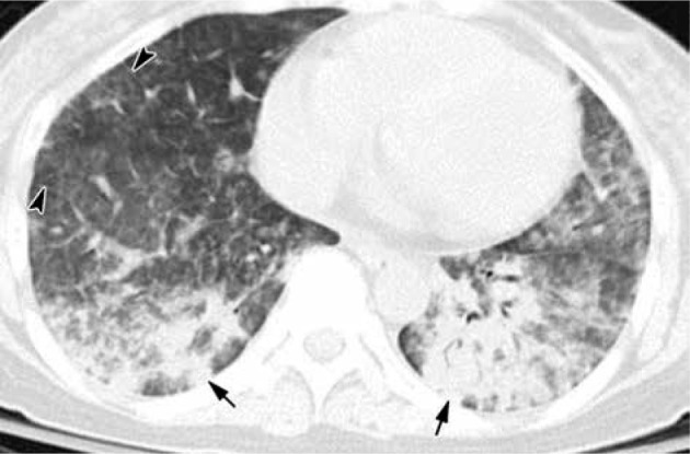
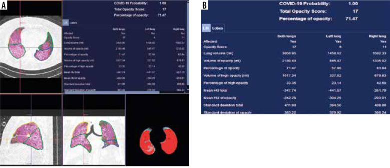
Chest computed tomography (CT) is proven to have high sensitivity in COVID-19 diagnosis. It is available in most emergency wards, and in contrast to polymerase chain reaction (PCR) it can be obtained in several minutes. However, its imaging features change during the course of the disease and overlap with other viral pneumonias, including influenza pneumonia. In this brief analysis we review the recent literature about chest CT features, useful radiological scales, and COVID-19 differentiation with other viral infections.

Keywords: COVID-19; chest CT; ground glass opacities; viral pneumonia.


Conflict of interest statement

The authors report no conflict of interest.

Figures

Figure 1
Figure 1
Multifocal ground-glass opacities
Figure 2
Figure 2
Air space consolidation. The image shows also ground-glass opacities, crazy paving, bronchovascular thickening, and vascular dilatation
Figure 3
Figure 3
Crazy paving appearance. The image shows also bronchovascular thickening, vascular dilatation, traction bronchiectasis, subpleural bands and architectural distortion, and air space consolidation
Figure 4
Figure 4
Reversed halo sign
Figure 5
Figure 5
COVID 19: Bilateral ground-glass opacities, crazy paving appearance, vascular dilatation, subpleural bands and architectural distortion. The distribution of changes is bilateral and peripheral
Figure 6
Figure 6
Influenza A pneumonia: computed tomography – consolidation (arrows), diffuse ground-glass opacities, interlobular septal thickening (arrowheads). The distribution of changes is mainly in the lower lobes. The peripheral distribution of consolidation is not as strong as in COVID-19 pneumonia (image from Koo et al. [35])
Figure 7
Figure 7
DL-based CT Pneumonia Analysis Prototype (Siemens Healthinners, Erlangen, Germany)

Similar articles

References

    1. Onigbinde SO, Ojo AS, Fleary L, et al. . Chest computed tomography findings in COVID-19 and influenza: a narrative review. Biomed Res Int 2020; 2020: 6928368. - PMC - PubMed
    1. Byambasuren S, Paradowska-Stankiewicz I, Brydak LB. Epidemic influenza seasons from 2008 to 2018 in Poland: a focused review of virological characteristics. Adv Exp Med Biol 2020; 1251: 115-121. - PMC - PubMed
    1. Fang Y, Zhang H, Xie J, et al. . Sensitivity of chest CT for COVID-19: comparison to RT-PCR. Radiology 2020; 296: 115-117. - PMC - PubMed
    1. Ai T, Yang Z, Hou H, et al. . Correlation of chest CT and RT-PCR testing for coronavirus disease 2019 (COVID-19) in China: a report of 1014 cases. Radiology 2020; 296: 32-40. - PMC - PubMed
    1. Kim H, Hong H, Yoon SH. Diagnostic performance of CT and reverse transcriptase polymerase chain reaction for coronavirus disease 2019: a meta-analysis. Radiology 2020; 296: 145-155. - PMC - PubMed