

Prenatally diagnosed accessory scrotum: A case report and review of the literature on prenatal features
- PMID: 35059092
- PMCID: PMC8760351
- DOI: 10.1016/j.radcr.2021.12.033
Prenatally diagnosed accessory scrotum: A case report and review of the literature on prenatal features
Abstract
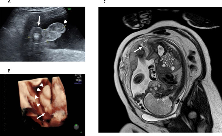
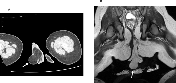
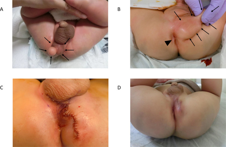
Accessory scrotum (AS) is rarely diagnosed antenatally, and its prenatal features remain unknown. Here, we report a case of a prenatally diagnosed accessory scrotum with perineal lipoma. A 33-year-old woman was referred to our hospital at 35 weeks of gestation to evaluate a mass in the fetal perineal region. Prenatal ultrasonography showed a 2.0 × 2.0 cm sized, echogenic, and circular mass located posterior to the left scrotum in a male fetus. Magnetic resonance imaging (MRI) showed a mass containing adipose tissue. A 6.5 cm elastic mass (AS and protruding lipoma) was observed in the perineal region, and surgical excision was performed at 8 months of age. Histological examination confirmed the diagnosis of AS with perineal lipoma, and there was no recurrence at follow-up. The typical prenatal presentation of AS was a circular perineal mass located posterior to the normal scrotum and was associated with perineal lipoma. The prenatal detection of AS was feasible with careful observation via ultrasonography, and prenatal MRI was useful in characterizing perineal tumors and evaluating associated anomalies.
Keywords: AS, Accessory scrotum; Accessory scrotum; CT, Computed tomography; MRI, Magnetic resonance imaging; Perineal lipoma; Prenatal diagnosis; US, Ultrasonography.
© 2021 The Authors. Published by Elsevier Inc. on behalf of University of Washington.
Conflict of interest statement
None.
Figures
Similar articles
-
Accessory scrotum with perineal lipoma diagnosed prenatally: case report and review of the literature.Nagoya J Med Sci. 2015 Aug;77(3):501-6. Nagoya J Med Sci. 2015. PMID: 26412897 Free PMC article.
-
Accessory scrotum attached to a perineal lipoma in an adult male.Int J Urol. 2003 Sep;10(9):501-3; discussion 504. doi: 10.1046/j.1442-2042.2003.00660.x. Int J Urol. 2003. PMID: 12941131
-
Congenital perianal lipoma: a case report and review of the literature.Surg Case Rep. 2019 Dec 16;5(1):199. doi: 10.1186/s40792-019-0753-z. Surg Case Rep. 2019. PMID: 31845000 Free PMC article.
-
[Congenital perineal lipoma with accessory scrotum: a case report].Hinyokika Kiyo. 1991 Jun;37(6):647-9. Hinyokika Kiyo. 1991. PMID: 1892016 Review. Japanese.
-
[Accessory scrotum with lipoma: a case report].Hinyokika Kiyo. 1996 Mar;42(3):233-6. Hinyokika Kiyo. 1996. PMID: 8619396 Review. Japanese.
Cited by
-
Accessory Scrotum - Truly an Appendage?J Indian Assoc Pediatr Surg. 2024 Jul-Aug;29(4):390-391. doi: 10.4103/jiaps.jiaps_54_24. Epub 2024 Jul 6. J Indian Assoc Pediatr Surg. 2024. PMID: 39149430 Free PMC article.
-
A case of dumbbell-shaped accessory scrotum with concomitant lipoma.Surg Case Rep. 2024 May 1;10(1):106. doi: 10.1186/s40792-024-01906-w. Surg Case Rep. 2024. PMID: 38691310 Free PMC article.
References
-
- Budhiraja S., Pandit S.K. Accessory Scrotum. Urol Int. 2000;63:210–211. - PubMed
Publication types
LinkOut - more resources
Full Text Sources
Research Materials
