Review
. 2021 Nov 25;9(Pt 1):11-20.
doi: 10.1107/S205225252101071X. eCollection 2022 Jan 1.

Using crystallography tools to improve vaccine formulations

Affiliations
Review

Using crystallography tools to improve vaccine formulations

Márcia Carvalho de Abreu Fantini et al. IUCrJ. .

Abstract

This article summarizes developments attained in oral vaccine formulations based on the encapsulation of antigen proteins inside porous silica matrices. These vaccine vehicles show great efficacy in protecting the proteins from the harsh acidic stomach medium, allowing the Peyer's patches in the small intestine to be reached and consequently enhancing immunity. Focusing on the pioneering research conducted at the Butantan Institute in Brazil, the optimization of the antigen encapsulation yield is reported, as well as their distribution inside the meso- and macroporous network of the porous silica. As the development of vaccines requires proper inclusion of antigens in the antibody cells, X-ray crystallography is one of the most commonly used techniques to unveil the structure of antibody-combining sites with protein antigens. Thus structural characterization and modelling of pure antigen structures, showing different dimensions, as well as their complexes, such as silica with encapsulated hepatitis B virus-like particles and diphtheria anatoxin, were performed using small-angle X-ray scattering, X-ray absorption spectroscopy, X-ray phase contrast tomography, and neutron and X-ray imaging. By combining crystallography with dynamic light scattering and transmission electron microscopy, a clearer picture of the proposed vaccine complexes is shown. Additionally, the stability of the immunogenic complex at different pH values and temperatures was checked and the efficacy of the proposed oral immunogenic complex was demonstrated. The latter was obtained by comparing the antibodies in mice with variable high and low antibody responses.

Keywords: SAXS; XAS; imaging; oral vaccines; porous silica.


Figures

Figure 1
Figure 1
Sketch of SBA-15 showing the macropores in the 20 µm particle, the 2 µm rod-shaped subunit and the 10 nm hexagonal-ordered mesopores [reproduced from the work by Rasmussen et al. (2017 ▸)].
Figure 2
Figure 2
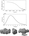
dANA SAXS results in PBS together with its corresponding PDF, p(r) and particle form obtained from a simulation model [reproduced from the work by Rasmussen et al. (2017 ▸)].
Figure 3
Figure 3
TEM image of dANA inside SBA-15 (L2) mesopores [reproduced from the work by Rasmussen et al. (2021 ▸)].
Figure 4
Figure 4
SAXS data for SBA-15 (L1) in PBS solution averaged over 1 h of measurement (points) and the corresponding fitting (continuous line).
Figure 5
Figure 5
SAXS data from the release of HBsAg from SBA-15 in PBS solution, pH 7.4. Changes in the scattering curve can be attributed to the release of HBsAg.
Figure 6
Figure 6
3D visualization of different agglomeration islands of HBsAg in the SBA-15 1:2 sample. Each agglomeration is shown using different colours for clarity.
Figure 7
Figure 7
(a) X-ray tomogram of SBA-15 1:1 showing clusters with different morphologies than observed for smaller ratios of HBsAg to SBA-15, obtained using the X-ray mCT setup. (b) Neutron tomogram of the SBA-15 1:1 sample showing clusters assigned to HBsAg with the same shape observed with X-rays, obtained with the CONRAD instrument. (c) 3D visualization of HBsAg aggregates in the SBA-15 1:1 sample. Each aggregate is shown with a different colour. These aggregates have a plate shape which is not observed in any other SBA-15 to HBsAg ratios. (d) 3D visualization of the SBA-15 particle using ID16B setup. The silica rods and some larger macropores can be observed, as indicated by the arrow (white region).
Figure 8
Figure 8
(a) STXM image of a particle from the 1:1 sample with 287.1 eV photon energy, just above the C=O bond peak showing differences of attenuation between the contamination around the particle and HBsAg. (b) STXM images of a particle from the 1:10 sample with 288.1 eV photon energy in the range attributed to C—H and C—N bonds.
Figure 9
Figure 9
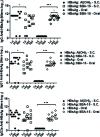
Anti-HBsAg serum IgG and subclass titers detected by ELISA in BALB/c mice immunized with HBsAg, encapsulated/adsorbed in mesoporous SBA-15 or adsorbed in Al(OH)3 by subcutaneous (SC) or oral (OR) routes. The titers were detected 7, 14 and 30 days after the booster. A group of animals immunized with rHBsAg was used as a reference for the unpaired Student t test analysis, *p < 0.05; ⋯ p < 0.001. Antibody titers at days 7, 14 and 30 after the booster were also used as a reference for the unpaired Student t test analysis, °p < 0.05 [reproduced from the work by Scaramuzzi et al., 2011 ▸)].
Figure 10
Figure 10
Anti-diphtheria serum IgG titers detected by ELISA in BALB/c mice immunized with dANA encapsulated/adsorbed in mesoporous SBA-15 or adsorbed in Al(OH)3 by subcutaneous (SC) or oral (OR) routes. The titers were detected 7 (grey columns) and 19 (black columns) days after immunization. The statistical analyses were performed with the one-way ANOVA test (*P < 0.05; ***P < 0.005) [reproduced from the work by Rasmussen et al. (2021 ▸)].

References

    1. Calderón, M. & Sosnik, A. (2015). Biotechnol. Adv. 33, 1277–1278. - PubMed
    1. Carvalho, L. V., Ruiz, R. D., Scaramuzzi, K., Marengo, E. B., Matos, J. R., Tambourgi, D. V., Fantini, M. C. A. & Sant’Anna, O. A. (2010). Vaccine, 28, 7829–7836. - PubMed
    1. Chen, L. J., Liu, J., Zhang, Y. L., Zhang, G. L., Kang, Y. Y., Chen, A. J., Feng, X. & Shao, L. Q. (2018). Nanomedicine, 13, 1939–1962. - PubMed
    1. Coria, L. M., Risso, G. S., Guaimas, F. F., Ferrero, M. C., Bruno, L., Pasquevich, K. A. & Cassataro, J. (2019). J. Controlled Release, 293, 158–171. - PMC - PubMed
    1. Dobrovolskaia, M. A., Germolec, D. R. & Weaver, J. L. (2009). Nat. Nanotech. 4, 411–414. - PubMed