Review
. 2021 Nov 18;4(2):100397.
doi: 10.1016/j.jhepr.2021.100397. eCollection 2022 Feb.

Transforming growth factor β latency: A mechanism of cytokine storage and signalling regulation in liver homeostasis and disease

Affiliations
Review

Transforming growth factor β latency: A mechanism of cytokine storage and signalling regulation in liver homeostasis and disease

Yujia Li et al. JHEP Rep. .

Abstract

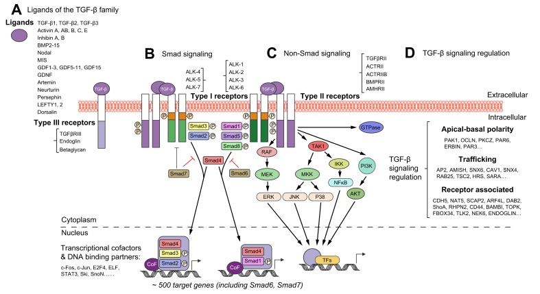
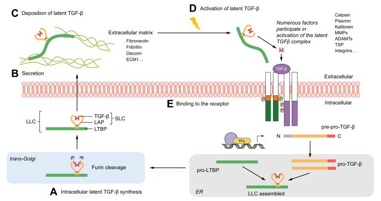
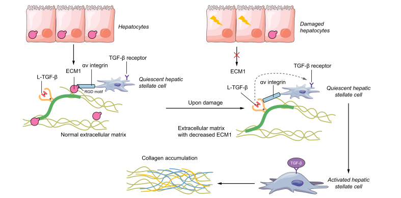
Transforming growth factor-β (TGF-β) is a potent effector in the liver, which is involved in a plethora of processes initiated upon liver injury. TGF-β affects parenchymal, non-parenchymal, and inflammatory cells in a highly context-dependent manner. Its bioavailability is critical for a fast response to various insults. In the liver - and probably in other organs - this is made possible by the deposition of a large portion of TGF-β in the extracellular matrix as an inactivated precursor form termed latent TGF-β (L-TGF-β). Several matrisomal proteins participate in matrix deposition, latent complex stabilisation, and activation of L-TGF-β. Extracellular matrix protein 1 (ECM1) was recently identified as a critical factor in maintaining the latency of deposited L-TGF-β in the healthy liver. Indeed, its depletion causes spontaneous TGF-β signalling activation with deleterious effects on liver architecture and function. This review article presents the current knowledge on intracellular L-TGF-β complex formation, secretion, matrix deposition, and activation and describes the proteins and processes involved. Further, we emphasise the therapeutic potential of toning down L-TGF-β activation in liver fibrosis and liver cancer.

Keywords: BMDCs, bone marrow-derived dendritic cells; BMPs, bone morphogenetic proteins; Co-Smad, co-mediator Smad; ECM, extracellular matrix; ECM1; ECM1, extracellular matrix protein 1; HCC, hepatocellular carcinoma; HSCs, hepatic stellate cells; I-Smad, inhibitory Smad; L-TGF-β, latent transforming growth factor β; LAP, latency-associated peptide; LLC, large latent complex; LTBP, latent TGF-β binding protein; Latent TGF-β; Liver disease; MMP, matrix metalloproteinase; PAI-1, plasminogen activator inhibitor-1; R-Smad, receptor-regulated Smad; RGD, arginine-glycine-aspartic acid; ROS, reactive oxygen species; SLC, small latent complex; TGF-β activation; TGF-β signalling; TGF-β, transforming growth factor β; TIMP-1, tissue inhibitor of metalloproteinase-1; TSP, thrombospondin; cFn, cellular fibronectin; cRGD, cyclic arginine-glycine-aspartic acid peptide; pFn, plasma fibronectin; rECM1, recombinant ECM1 protein; α-SMA, alpha-smooth muscle actin.


Conflict of interest statement

The authors declare that they have no conflict of interest. Please refer to the accompanying ICMJE disclosure forms for further details.

Figures

Fig. 1
Fig. 1
The TGF-β signalling pathway. The scheme comprises different ligands and receptors of the TGF-β family, canonical Smad and non-canonical signalling pathways, and the network of regulatory interactions at multiple levels from the cell surface to the nucleus. (A) TGF-β family members, including TGF-β1, TGF-β2, TGF-β3, activins, inhibins, BMPs, nodal, MIS, and GDFs, initiate multiple cellular responses and cell fate decisions. (B) Secreted as protein ligands in latent and inactive forms, upon activation they may be presented by type III receptors (TGFβRIII or Endoglin) lacking kinase activity to the signalling receptor complex, consisting of heteromeric type II and type I receptors. Ligand binding to transmembrane type II receptors (e.g. TGFβRII, ACTRII, BMPRII) leads to autophosphorylation, recruitment and phosphorylation of type I receptor kinases (e.g. ALK-4, ALK-5, ALK-7 and ALK-1, ALK-2), thus forming a ligand-receptor complex to phosphorylate and activate the canonical Smad signalling pathway. Activated R-Smads (Smad2, Smad3 and Smad1, Smad5, Smad8, can be phosphorylated by ALK-4, ALK-5, ALK-7 and ALK-1, ALK-2, respectively) form hetero-oligomers with Co-Smads (Smad4), and the heteromeric complexes then translocate into the nucleus, where, in conjunction with variant co-factors and DNA-binding partners, such as c-Fos, c-Jun, ELF, or Ski, SnoN, and CEBP, they modulate chromatin decondensation and regulate target gene transcription. The I-Smads Smad6 and Smad7, which are direct transcriptional targets of Smad signalling, form crucial negative feedback loops to TGF-β signalling. (C) Non-Smad TGFβR signalling pathways are also frequently involved in regulating intracellular TGF-β signalling. TAK1 plays a central role, potentially coordinating the activation of MAPK signalling, including JNK, p38 MAPK, and NF-κB signalling. MEK-ERK, PI3K-AKT and some Rho family members like GTPases were also identified in the large TGF-β signalling crosstalk. (D) TGF-β signalling regulation occurs at multiple levels in extracellular and intracellular spaces. Thereby, the core components of the TGF-β-Smad signalling pathway are embedded within a huge network of hierarchical protein-protein interactions, leading to regulatory nodes at multiple levels. BMPs, bone morphogenetic proteins; Co-Smad, co-mediator Smad; I-Smad, inhibitory Smad; GDFs, growth differentiation factors; MIS, Müllerian-inhibiting substance; R-Smad, receptor-regulated Smad; TGF-β, transforming growth factor β.
Fig. 2
Fig. 2
Graphical summary of TGF-β protein production, secretion, matrix deposition, and maturation. (A) (starting at the bottom) After translation of pre-pro-TGF-β, the N-terminal signal peptide is cleaved to yield pro-TGF-β which translocates into the ER (lower right), where pro-TGF-β molecules dimerise and form disulphide bonds to LTBP, forming a ternary complex. The TGF-β dimer is cleaved from its pro-peptide in the trans-Golgi (lower left) but remains strongly associated with the LAP via non-covalent interactions. TGF-β and LAP form the SLC, which together with LTBP comprise the LLC. (B) Once secreted (middle left), the latent TGF-β is deposited into the extracellular matrix (C) (upper left), thanks to the high affinity of LTBP to various matrix components, such as fibronectin, fibrillin, and decorin. (D) (upper right) Numerous factors, like physiological proteins including proteases, thrombospondin and integrins, physical conditions and chemicals, including ROS and lactic acid, participate in activation of latent TGF-β, leading to the release of the mature TGF-β signalling ligand. The active ligand then binds to the cell surface receptors (E) (middle right) to regulate a wide variety of downstream signalling pathways and cellular responses. ER, endoplasmic reticulum; LAP, latency-associated peptide; LLC, large latent complex; LTBP, latent TGF-β binding protein; ROS, reactive oxygen species; SLC, small latent complex; TGF-β, transforming growth factor β.
Fig. 3
Fig. 3
Scheme showing how ECM1 sequesters the latent TGF-β from integrin activation. We recently found that ECM1, a glycoprotein in the extracellular matrix of the liver, is critical in maintaining normal architecture and physiological homeostasis. Under normal conditions, ECM1 binds to the RGD motif, which commonly exists in the LAP and most integrins, thus inhibiting the activation between integrins and latent TGF-β. The presence of ECM1 is critical to keep extracellular matrix-deposited TGF-β in a latent and quiescent form, preventing HSC activation and liver fibrosis. Conversely, depleting the liver of ECM1 results in spontaneous release of active TGF-β ligand from the latent complex, leading to strongly enhanced TGF-β signalling, HSC activation, collagen accumulation, ultimately promoting spontaneous architectural disturbances and severe liver fibrosis. ECM1, extracellular matrix protein 1; HSC, hepatic stellate cell; LAP, latency-associated peptide; L-TGF-β, latent TGF-β; RGD, arginine-glycine-aspartic acid; TGF-β, transforming growth factor β.

Similar articles

Cited by

References

    1. Roberts A.B., Anzano M.A., Lamb L.C., Smith J.M., Sporn M.B. New class of transforming growth factors potentiated by epidermal growth factor: isolation from non-neoplastic tissues. Proc Natl Acad Sci U S A. 1981;78:5339–5343. - PMC - PubMed
    1. Taipale J., Saharinen J., Keski-Oja J. Extracellular matrix-associated transforming growth factor-beta: role in cancer cell growth and invasion. Adv Cancer Res. 1998;75:87–134. - PubMed
    1. Laiho M., Keski-Oja J. Transforming growth factors-beta as regulators of cellular growth and phenotype. Crit Rev Oncog. 1992;3:1–26. - PubMed
    1. Morikawa M., Derynck R., Miyazono K. TGF-beta and the TGF-beta family: context-dependent roles in cell and tissue physiology. Cold Spring Harb Perspect Biol. 2016;8 - PMC - PubMed
    1. Shi Y., Massague J. Mechanisms of TGF-beta signaling from cell membrane to the nucleus. Cell. 2003;113:685–700. - PubMed