

Circulating cardiomyocyte-derived extracellular vesicles reflect cardiac injury during systemic inflammatory response syndrome in mice
- PMID: 35059851
- PMCID: PMC8776681
- DOI: 10.1007/s00018-021-04125-w
Circulating cardiomyocyte-derived extracellular vesicles reflect cardiac injury during systemic inflammatory response syndrome in mice
Abstract
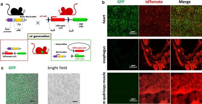
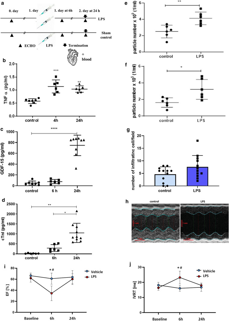
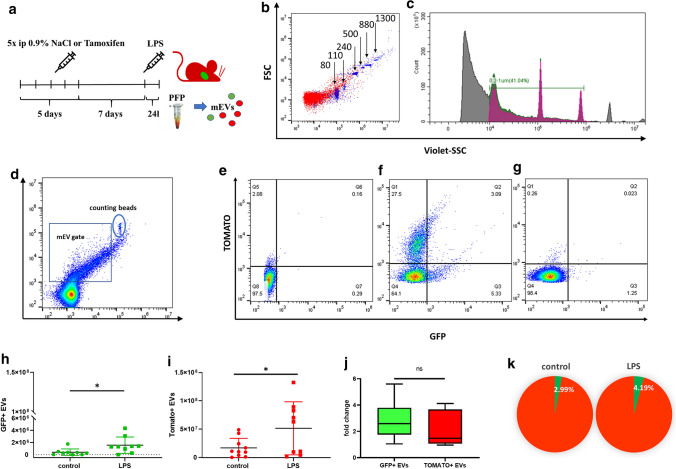
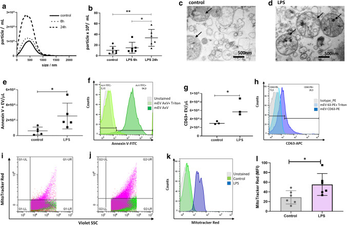
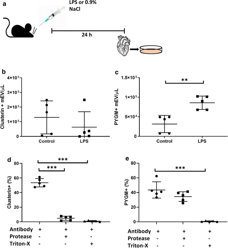
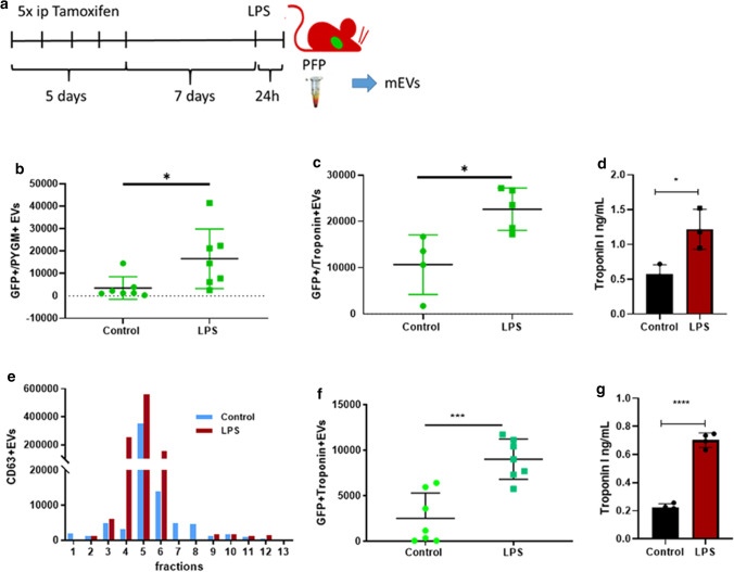
The release of extracellular vesicles (EVs) is increased under cellular stress and cardiomyocyte damaging conditions. However, whether the cardiomyocyte-derived EVs eventually reach the systemic circulation and whether their number in the bloodstream reflects cardiac injury, remains unknown. Wild type C57B/6 and conditional transgenic mice expressing green fluorescent protein (GFP) by cardiomyocytes were studied in lipopolysaccharide (LPS)-induced systemic inflammatory response syndrome (SIRS). EVs were separated both from platelet-free plasma and from the conditioned medium of isolated cardiomyocytes of the left ventricular wall. Size distribution and concentration of the released particles were determined by Nanoparticle Tracking Analysis. The presence of GFP + cardiomyocyte-derived circulating EVs was monitored by flow cytometry and cardiac function was assessed by echocardiography. In LPS-treated mice, systemic inflammation and the consequent cardiomyopathy were verified by elevated plasma levels of TNFα, GDF-15, and cardiac troponin I, and by a decrease in the ejection fraction. Furthermore, we demonstrated elevated levels of circulating small- and medium-sized EVs in the LPS-injected mice. Importantly, we detected GFP+ cardiomyocyte-derived EVs in the circulation of control mice, and the number of these circulating GFP+ vesicles increased significantly upon intraperitoneal LPS administration (P = 0.029). The cardiomyocyte-derived GFP+ EVs were also positive for intravesicular troponin I (cTnI) and muscle-associated glycogen phosphorylase (PYGM). This is the first direct demonstration that cardiomyocyte-derived EVs are present in the circulation and that the increased number of cardiac-derived EVs in the blood reflects cardiac injury in LPS-induced systemic inflammation (SIRS).
Keywords: Cardiomyocyte; Cardiomyopathy; Extracellular vesicles; Inducible transgenic mice; SIRS.
© 2022. The Author(s).
Conflict of interest statement
EIB is a member of the Advisory Board of Sphere Gene Therapeutics Inc. (Boston, MA, US). PF is the founder and CEO of Pharmahungary Group, a group of R&D companies.
Figures
References
-
- Hegyesi H, Sándor N, Sáfrány G, Lovas V, Kovács Á, Takács A, Kőhidai L, Turiák L, Kittel Á, Pálóczi K, Bertók L, Buzás EI. Radio-detoxified LPS alters bone marrow-derived extracellular vesicles and endothelial progenitor cells. Stem Cell Res Ther. 2019;10:313. doi: 10.1186/s13287-019-1417-4. - DOI - PMC - PubMed
-
- Théry C, Witwer KW, Aikawa E, Alcaraz MJ, Anderson JD, Andriantsitohaina R, Antoniou A, Arab T, Archer F, Atkin-Smith GK, Ayre DC, Bach JM, Bachurski D, Baharvand H, Balaj L, Baldacchino S, Bauer NN, Baxter AA, Bebawy M, Beckham C, Bedina Zavec A, Benmoussa A, Berardi AC, Bergese P, Bielska E, Blenkiron C, Bobis-Wozowicz S, Boilard E, Boireau W, Bongiovanni A, Borràs FE, Bosch S, Boulanger CM, Breakefield X, Breglio AM, Brennan MÁ, Brigstock DR, Brisson A, Broekman ML, Bromberg JF, Bryl-Górecka P, Buch S, Buck AH, Burger D, Busatto S, Buschmann D, Bussolati B, Buzás EI, Byrd JB, Camussi G, Carter DR, Caruso S, Chamley LW, Chang YT, Chen C, Chen S, Cheng L, Chin AR, Clayton A, Clerici SP, Cocks A, Cocucci E, Coffey RJ, Cordeiro-da-Silva A, Couch Y, Coumans FA, Coyle B, Crescitelli R, Criado MF, D’Souza-Schorey C, Das S, Datta Chaudhuri A, de Candia P, De Santana EF, De Wever O, De Portillo HA, Demaret T, Deville S, Devitt A, Dhondt B, Di Vizio D, Dieterich LC, Dolo V, Dominguez-Rubio AP, Dominici M, Dourado MR, Driedonks TA, Duarte FV, Duncan HM, Eichenberger RM, Ekström K, El-Andaloussi S, Elie-Caille C, Erdbrügger U, Falcón-Pérez JM, Fatima F, Fish JE, Flores-Bellver M, Försönits A, Frelet-Barrand A, Fricke F, Fuhrmann G, Gabrielsson S, Gámez-Valero A, Gardiner C, Gärtner K, Gaudin R, Gho YS, Giebel B, Gilbert C, Gimona M, Giusti I, Goberdhan DC, Görgens A, Gorski SM, Greening DW, Gross JC, Gualerzi A, Gupta GN, Gustafson D, Handberg A, Haraszti RA, Harrison P, Hegyesi H, Hendrix A, Hill AF, Hochberg FH, Hoffmann KF, Holder B, Holthofer H, Hosseinkhani B, Hu G, Huang Y, Huber V, Hunt S, Ibrahim AG, Ikezu T, Inal JM, Isin M, Ivanova A, Jackson HK, Jacobsen S, Jay SM, Jayachandran M, Jenster G, Jiang L, Johnson SM, Jones JC, Jong A, Jovanovic-Talisman T, Jung S, Kalluri R, Kano SI, Kaur S, Kawamura Y, Keller ET, Khamari D, Khomyakova E, Khvorova A, Kierulf P, Kim KP, Kislinger T, Klingeborn M, Klinke DJ, Kornek M, Kosanović MM, Kovács ÁF, Krämer-Albers EM, Krasemann S, Krause M, Kurochkin IV, Kusuma GD, Kuypers S, Laitinen S, Langevin SM, Languino LR, Lannigan J, Lässer C, Laurent LC, Lavieu G, Lázaro-Ibáñez E, Le-Lay S, Lee MS, Lee YXF, Lemos DS, Lenassi M, Leszczynska A, Li IT, Liao K, Libregts SF, Ligeti E, Lim R, Lim SK, Linē A, Linnemannstöns K, Llorente A, Lombard CA, Lorenowicz MJ, Lörincz ÁM, Lötvall J, Lovett J, Lowry MC, Loyer X, Lu Q, Lukomska B, Lunavat TR, Maas SL, Malhi H, Marcilla A, Mariani J, Mariscal J, Martens-Uzunova ES, Martin-Jaular L, Martinez MC, Martins VR, Mathieu M, Mathivanan S, Maugeri M, McGinnis LK, McVey MJ, Meckes DG, Meehan KL, Mertens I, Minciacchi VR, Möller A, Møller Jørgensen M, Morales-Kastresana A, Morhayim J, Mullier F, Muraca M, Musante L, Mussack V, Muth DC, Myburgh KH, Najrana T, Nawaz M, Nazarenko I, Nejsum P, Neri C, Neri T, Nieuwland R, Nimrichter L, Nolan JP, Nolte-‘t Hoen EN, Noren-Hooten N, O'Driscoll L, O'Grady T, O'Loghlen A, Ochiya T, Olivier M, Ortiz A, Ortiz LA, Osteikoetxea X, Østergaard O, Ostrowski M, Park J, Pegtel DM, Peinado H, Perut F, Pfaffl MW, Phinney DG, Pieters BC, Pink RC, Pisetsky DS, Pogge von Strandmann E, Polakovicova I, Poon IK, Powell BH, Prada I, Pulliam L, Quesenberry P, Radeghieri A, Raffai RL, Raimondo S, Rak J, Ramirez MI, Raposo G, Rayyan MS, Regev-Rudzki N, Ricklefs FL, Robbins PD, Roberts DD, Rodrigues SC, Rohde E, Rome S, Rouschop KM, Rughetti A, Russell AE, Saá P, Sahoo S, Salas-Huenuleo E, Sánchez C, Saugstad JA, Saul MJ, Schiffelers RM, Schneider R, Schøyen TH, Scott A, Shahaj E, Sharma S, Shatnyeva O, Shekari F, Shelke GV, Shetty AK, Shiba K, Siljander PR, Silva AM, Skowronek A, Snyder OL, Soares RP, Sódar BW, Soekmadji C, Sotillo J, Stahl PD, Stoorvogel W, Stott SL, Strasser EF, Swift S, Tahara H, Tewari M, Timms K, Tiwari S, Tixeira R, Tkach M, Toh WS, Tomasini R, Torrecilhas AC, Tosar JP, Toxavidis V, Urbanelli L, Vader P, van Balkom BW, van der Grein SG, Van Deun J, van Herwijnen MJ, Van Keuren-Jensen K, van Niel G, van Royen ME, van Wijnen AJ, Vasconcelos MH, Vechetti IJ, Jr, Veit TD, Vella LJ, Velot É, Verweij FJ, Vestad B, Viñas JL, Visnovitz T, Vukman KV, Wahlgren J, Watson DC, Wauben MH, Weaver A, Webber JP, Weber V, Wehman AM, Weiss DJ, Welsh JA, Wendt S, Wheelock AM, Wiener Z, Witte L, Wolfram J, Xagorari A, Xander P, Xu J, Yan X, Yáñez-Mó M, Yin H, Yuana Y, Zappulli V, Zarubova J, Žėkas V, Zhang JY, Zhao Z, Zheng L, Zheutlin AR, Zickler AM, Zimmermann P, Zivkovic AM, Zocco D, Zuba-Surma EK. Minimal information for studies of extracellular vesicles 2018 (MISEV2018): a position statement of the International Society for extracellular vesicles and update of the MISEV2014 guidelines. J Extracell Vesicles. 2018;7:1530. doi: 10.1080/20013078.2018.1535750. - DOI - PMC - PubMed
MeSH terms
Substances
Grants and funding
- NVKP_16-1-2016-0017/nemzeti kutatási, fejlesztési és innovaciós alap
- VEKOP-2.3.2-16-2016-000002/nemzeti kutatási, fejlesztési és innovaciós alap
- VEKOP-2.3.3-15-2017-00016/nemzeti kutatási, fejlesztési és innovaciós alap
- Higher Education Excellence Program (FIKP) Therapeutic Thematic Programme/nemzeti kutatási, fejlesztési és innovaciós alap
- TKP2020-NKA-26/nemzeti kutatási, fejlesztési és innovaciós alap
LinkOut - more resources
Full Text Sources
Research Materials
