. 2023 Jan;43(1):367-380.
doi: 10.1007/s10571-022-01190-y. Epub 2022 Jan 21.

Modulating Expression of Endogenous Interleukin 1 Beta in the Acute Phase of the Pilocarpine Model of Epilepsy May Change Animal Survival

Affiliations

Modulating Expression of Endogenous Interleukin 1 Beta in the Acute Phase of the Pilocarpine Model of Epilepsy May Change Animal Survival

V D B Pascoal et al. Cell Mol Neurobiol. 2023 Jan.

Abstract

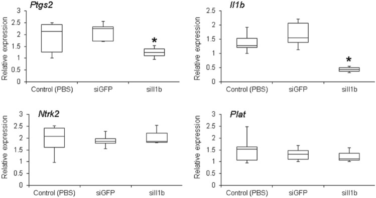
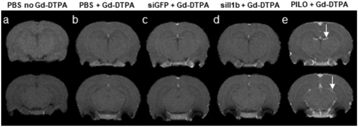
The pilocarpine-induced (PILO) model has helped elucidate the electrophysiological and molecular aspects related to mesial temporal lobe epilepsy. It has been suggested that the extensive cell death and edema observed in the brains of these animals could be induced by increased inflammatory responses, such as the rapid release of the inflammatory cytokine interleukin 1 beta (Il1b). In this study, we investigate the role of endogenous Il1b in the acute phase of the PILO model. Our aim is twofold. First, we want to determine whether it is feasible to silence Il1b in the central nervous system using a non-invasive procedure. Second, we aim to investigate the effect of silencing endogenous Il1b and its antagonist, Il1rn.We used RNA interference applied non-invasively to knockdown Il1b and its endogenous antagonist Il1rn. We found that knocking down Il1b prior to pilocarpine injection increased the mortality rate of treated animals. Furthermore, we observed that, when exposing the animals to more Il1b by silencing its endogenous antagonist Il1rn, there was a better response to status epilepticus with decreased animal mortality in the acute phase of the PILO model. Thus, we show the feasibility of using a novel, less invasive approach to study genes involved in the inflammatory response in the central nervous system. Furthermore, our results provide suggestive evidence that modulating endogenous Il1b improves animal survival in the acute phase of the PILO model and may have effects that extend into the chronic phase.

Keywords: Animal model; Mesial temporal lobe epilepsy; Neuroinflammation; RNA interference in vivo.


Conflict of interest statement

None of the authors have any conflict of interest to disclose.

Figures

Fig. 1
Fig. 1
Gene silencing effect of siIl1b over time. Animals were injected twice (8-h interval between injections) with 25 μg of siIl1b in the tail vein. Gene expression was quantified in the whole brain by real-time polymerase chain reaction (qPCR). Control animals were injected with phosphate-buffered saline (PBS) or siGFP (data not shown). All groups were composed of three animals. Asterisk indicates significant gene silencing (Kruskal–Wallis test, p < 0.05). p.i., post-injection
Fig. 2
Fig. 2
Assessment of RNA interference specificity. Relative gene expression quantification [real-time polymerase chain reaction (qPCR)] of Ptgs2, Il1b, Ntrk2, and Plat in animals that received two injections of 25 μg of siRNA (8-h interval between injections). Brains were collected and RNA was extracted 72 h after the first injection. As expected, phosphate-buffered saline (PBS) alone or siGFP did not affect the expression of any of the assessed genes. By contrast, siIl1b led to the silencing of both Il1b and Ptgs2 (which are in the same biological pathway as Il1b), whereas Ntrk2 and Plat (which are not biologically related to Il1b) were not affected. Each experiment was performed in the whole brain and extracted from five animals. Asterisk indicates significant gene silencing (Kruskal–Wallis test, p < 0.05)
Fig. 3
Fig. 3
Representative magnetic resonance images of animals used in our experiments with RNA interference. We selected two slices near the middle of the cerebrum and another one in the hippocampal region: a images from an animal that was injected with PBS but not paramagnetic contrast (gadolinium diethylenetriaminepentacetate [Gd-DTPA]); b animals injected with phosphate-buffered saline (PBS, control) followed by Gd-DTPA; c animals injected with siGFP::RVG9R followed by Gd-DTPA; d animals injected with siIl1b::RVG9R followed by Gd-DTPA; and e animals injected with Gd-DTPA 2 h after status epilepticus induced by pilocarpine (PILO) injection (positive control). All groups were composed of three animals. The white arrows indicate the presence of Gd-DTPA (intensified signal) within the brain as a marker of BBB damage
Fig. 4
Fig. 4
Il1b and Il1rn gene expression profiles over time in the brain after pilocarpine injection. The expression profiles were generated by using real-time polymerase chain reaction (qPCR) of both genes 1, 3, 6, and 24 h post-injection (p.i.) of pilocarpine and compared with control (no siIl1b or pilocarpine administration). We observed an increase in Il1b expression at 6 h p.i. All groups were composed of three animals. Asterisk indicates significant changes in gene expression (Kruskal–Wallis test, p < 0.05)
Fig. 5
Fig. 5
Phenotype assessment during the acute phase of the pilocarpine-induced epilepsy model. a The onset for the first seizure was 34 min (standard deviation [SD] = 12) in controls, 26 min (SD = 14) in animals pre-injected with siIl1b, and 49 min (SD = 16) in animals pre-injected with siIl1rn. b The onset of the status epilepticus (SE) was: 58 min (SD = 19) in controls, 46 min (SD = 25) in animals pre-injected with siIl1b, and 70 min (SD = 18) in animals pre-injected with siIl1rn. Single and double asterisk indicate a significant difference compared with the control group (analysis of variance with Tukey’s test, p < 0.05 and p < 0.01, respectively)
Fig. 6
Fig. 6
Analysis of gene expression 48 h after siIl1b or siIl1rn injection (no pilocarpine administration). Real-time polymerase chain reaction (qPCR) was used to determine the messenger RNA (mRNA) levels of Il1b, Il1rn, Nfkb, and Slc1a3 in animals injected with siIl1b or siIl1rn. Control animals were injected with phosphate-buffered saline (PBS). All groups were composed of five animals. Single and double asterisk indicate a significant difference in gene expression (Kruskal–Wallis test, p < 0.05 and p < 0.01, respectively)
Fig. 7
Fig. 7
Nissl staining of the hippocampal subfields for neuronal count (cells/µm2) of animals in the chronic phase of the pilocarpine model. Animals injected twice (8-h interval between injections) with 25 μg of siIl1rn in the tail vein 48 h before pilocarpine administration and survived after status epilepticus were kept under observation for 90 days. The images show the following: a hilus and DG, c CA1, and e CA3 regions of control animals injected with pilocarpine and PBS; b hilus and DG, d CA1, and f CA3 regions of animals pre-treated with of siIl1rn. We observed a discrete neuronal loss in the hilus, DG, CA1, and CA3 regions in animals pre-treated with siIl1rn (ANOVA and Tukey’s test, p < 0.01). Scale bar 180 µm

References

    1. Al Morshedy S, Elsaadany HF, Ibrahim HE et al (2017) Interleukin-1β and interleukin-1receptor antagonist polymorphisms in Egyptian children with febrile seizures: a case-control study. Medicine 96:e6370. 10.1097/MD.0000000000006370 - PMC - PubMed
    1. Allan SM, Tyrrell PJ, Rothwell NJ (2005) Interleukin-1 and neuronal injury. Nat Rev Immunol 5:629–640. 10.1038/nri1664 - PubMed
    1. Arida RM, Scorza FA, Peres CA, Cavalheiro EA (1999) The course of untreated seizures in the pilocarpine model of epilepsy. Epilepsy Res 34:99–107. 10.1016/s0920-1211(98)00092-8 - PubMed
    1. Arzimanoglou A, Hirsch E, Nehlig A et al (2002) Epilepsy and neuroprotection: an illustrated review. Epileptic Disord 4:173–182 - PubMed
    1. Avital A, Goshen I, Kamsler A et al (2003) Impaired interleukin-1 signaling is associated with deficits in hippocampal memory processes and neural plasticity. Hippocampus 13:826–834. 10.1002/hipo.10135 - PubMed

MeSH terms