. 2022 Jan 21;8(3):eabj6901.
doi: 10.1126/sciadv.abj6901. Epub 2022 Jan 21.

In vivo delivery of CRISPR-Cas9 using lipid nanoparticles enables antithrombin gene editing for sustainable hemophilia A and B therapy

Affiliations

In vivo delivery of CRISPR-Cas9 using lipid nanoparticles enables antithrombin gene editing for sustainable hemophilia A and B therapy

Jeong Pil Han et al. Sci Adv. .

Abstract

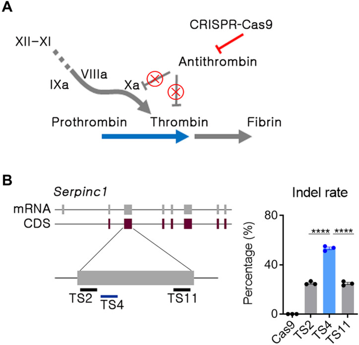
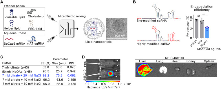
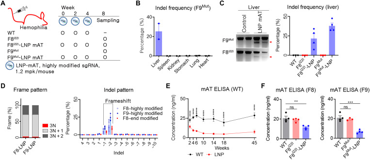
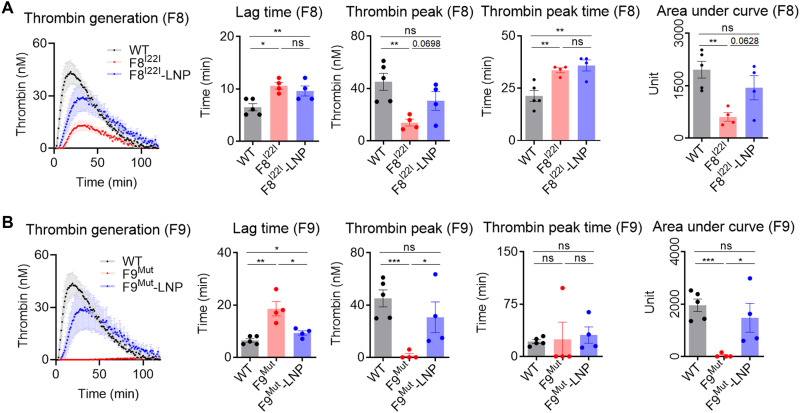
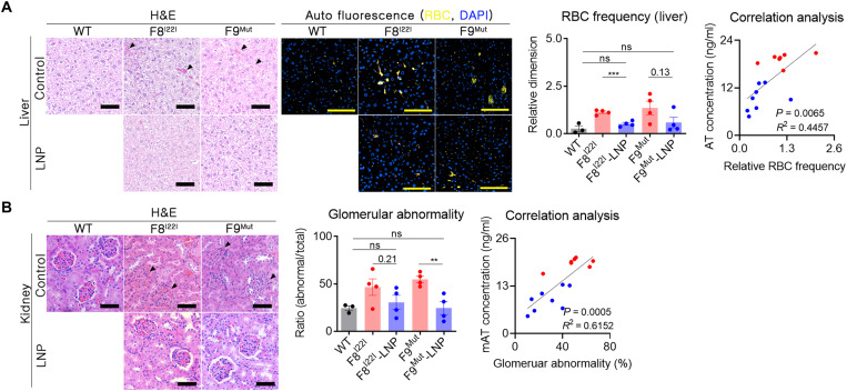
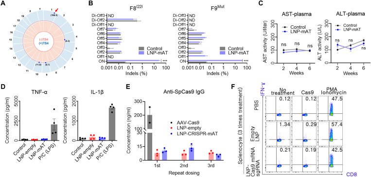
Hemophilia is a hereditary disease that remains incurable. Although innovative treatments such as gene therapy or bispecific antibody therapy have been introduced, substantial unmet needs still exist with respect to achieving long-lasting therapeutic effects and treatment options for inhibitor patients. Antithrombin (AT), an endogenous negative regulator of thrombin generation, is a potent genome editing target for sustainable treatment of patients with hemophilia A and B. In this study, we developed and optimized lipid nanoparticles (LNPs) to deliver Cas9 mRNA along with single guide RNA that targeted AT in the mouse liver. The LNP-mediated CRISPR-Cas9 delivery resulted in the inhibition of AT that led to improvement in thrombin generation. Bleeding-associated phenotypes were recovered in both hemophilia A and B mice. No active off-targets, liver-induced toxicity, and substantial anti-Cas9 immune responses were detected, indicating that the LNP-mediated CRISPR-Cas9 delivery was a safe and efficient approach for hemophilia therapy.


Figures

Fig. 1.
Fig. 1.. The AT locus was selected as the target gene for rebalancing.
(A) Strategy for CRISPR-Cas9–mediated in vivo Serpinc1 (encoding AT) gene editing. The red line and “X” symbols indicate inhibition of gene expression or its function. (B) Single guide RNAs (sgRNAs) were selected in the second exon of the Serpinc1 gene, and double-stranded breakage potential was evaluated by deep sequencing after transfection of C2C12 cells with an RNP formulation. Transfection and sequencing were conducted in triplicates, and each dot indicates the double-stranded breakage frequency from each experiment. TS, target site. Data were presented as mean ± SEM. ****P < 0.001.
Fig. 2.
Fig. 2.. LNPs for CRISPR-Cas9–mediated in vivo gene editing were prepared.
(A) Schematic image of LNP formulation using a microfluidic mixing system. (B) Encapsulation efficiency (EE) was analyzed between end- and highly modified sgRNA at 7 mM citrate buffer (n = 5). In the sgRNA structure, the asterisks and red font indicate the phosphorothioate bond and 2′-O-methyl ribonucleotide, respectively. Data are exhibited as means ± SEM. ***P < 0.001. (C) EE, size, and PDI of LNPs were analyzed with different buffer conditions. The condition selected in this study has been indicated in blue. (D) Biodistribution was analyzed by in vivo bioluminescence imaging. mFlucLNPs were formulated using the Luc mRNA (0.1 mpk dose) and injected intravenously into C57BL/6 mice. Three hours after the injection, luminescence signal was detected in the live mice and in their organs.
Fig. 3.
Fig. 3.. LNP-CRISPR-mAT induced a prolonged down-regulation of mAT expression.
(A) Brief schematic for the in vivo gene targeting using LNP-CRISPR-mAT. C57BL6 (B6, n = 4), B6.FVIII intron 22 inversion (F8I22I, n = 4 in each group), and B6.FIX knockout (F9Mut, n = 4 in each group) mice were used in this study. (B and C) Indel frequency was calculated by deep sequencing and confirmed using T7E1 analysis (number of each group, 2 or 4). Data are shown as means ± SEM. (D) Indel pattern was analyzed according to frameshift and indel size (number of each group, 3 or 4). Each dot indicates the percentage of each sized indel from the total sequencing result. (E) Prolonged AT down-regulation was screened by ELISA using B6 mice (n = 6 per group). Blood was collected from the tail vein using a sodium citrate–coated tube, and the plasma was subjected to mAT ELISA. ****P < 0.0001. (F) Blood mAT concentration was measured and compared between the LNP-CRISPR-mAT–treated and the control hemophilia mice group. Each dot indicates the mAT concentration of an individual mouse (number of each group, 3 or 4). Data are presented as means ± SEM. **P < 0.01 and ***P < 0.001; ns, nonsignificant.
Fig. 4.
Fig. 4.. In vivo mAT targeting rescued thrombogenesis.
(A and B) Plasma from F8I22I and F9Mut mice were collected from the inferior vena cava (control WT, n = 5; F8I22I, F8I22I-LNP, F9Mut, and F9Mut-LNP, n = 4). The thrombogenesis potential was calculated and analyzed for lag time, thrombin peak, thrombin peak time, and area under the curve. Each dot indicates data from one mouse and represents means ± SEM. *P < 0.05, **P < 0.01 and ***P < 0.001. Thrombogenesis in WT mice was assessed once and used for comparison with the F8I22I and F9Mut mice.
Fig. 5.
Fig. 5.. Enhanced thrombosis reduced spontaneous bleeding and secondary hemophilia complication.
(A) Paraffin-embedded liver tissues were prepared without perfusion to prevent the loss of evidence of spontaneous bleeding (WT, n = 3; F8I22I, F8I22I-LNP, F9Mut, and F9Mut-LNP, n = 4). RBCs in the interstitial liver tissue [marked by black triangles in the liver tissue using hematoxylin and eosin (H&E) staining] were measured by immunofluorescence staining by detecting the autofluorescence signal at 470 and 540 nm. The intensity of the coexpressions (yellow signals) were then calculated. Ten areas of the liver were randomly selected from each mouse, and each measured autofluorescence signal was subjected to the RBC frequency analysis. Each dot indicates a signal from one selected area. Correlation analysis was conducted using the mean yellow fluorescence values obtained from each mouse. Blue dot, LNP-CRISPR-mAT groups; red dot, control group. Scale bars, 100 μm. (B) The whole kidney was formalin fixed, and gross histology was examined using H&E staining. The number of abnormally shaped glomerular capsules (black triangles) was calculated from three randomly selected regions of the cortex (1 mm2) per mouse and analyzed. **P < 0.01 and ***P < 0.001.
Fig. 6.
Fig. 6.. Safety-related assessments of the LNP-CRISPR-Cas9.
(A) Genome-wide Circos plot for in vitro cleavage sites in the absence (pink) or presence (blue) of the TS4 sgRNA. Numbers in bracket: cleavage scores. Red arrow: on-target cleavage (B) Targeted deep sequencing results of the top seven homologous candidates (Offs) and the three candidates detected by the Digenome-seq analysis (Di-Offs) (n = 3). ND, not detected; ***P < 0.001. (C) Serum AST and ALT concentrations after injection with 1.2 mpk of LNP-CRISPR-mAT thrice to WT at an interval of 2 weeks (n = 6). (D) Serum TNF-α and IL-1β concentration after injection with 1.2 mpk of LNP or LNP-CRISPR-mAT thrice. Positive control group was injected 20 mpk of lipopolysaccharide (n = 4). (E) Serum anti-SpCas9 IgG concentration after repeated injection with LNP-CRISPR-mAT (n = 3). Mouse intravenously injected with AAV9-EFS-SpCas9 (5 × 10¹³ vg/kg) was also tested after 6 weeks of the treatment. The concentrations were calculated from the standard curve from ELISA (R2 = 0.989). (F) Representative flow cytometry plots illustrating IFN-γ expression in CD8+ T cells. IFN-γ expression in CD8+ T cells was evaluated after repeated injections of PBS, empty LNPs, and sgRNA/Cas9 mRNA–encapsulated LNPs to mice (n = 3). Detailed results are supplied in fig. S5.

References

    1. Peters R., Harris T., Advances and innovations in haemophilia treatment. Nat. Rev. Drug Discov. 17, 493–508 (2018). - PubMed
    1. Bulcha J. T., Wang Y., Ma H., Tai P. W. L., Gao G., Viral vector platforms within the gene therapy landscape. Signal Transduct. Target. Ther. 6, 53 (2021). - PMC - PubMed
    1. Valentino L. A., Cooper D. L., Goldstein B., Surgical experience with rFVIIa (NovoSeven) in congenital haemophilia A and B patients with inhibitors to factors VIII or IX. Haemophilia 17, 579–589 (2011). - PubMed
    1. van den Berg H. M., Fischer K., Carcao M., Chambost H., Kenet G., Kurnik K., Konigs C., Male C., Santagostino E., Ljung R.; PedNet Study Group , Timing of inhibitor development in more than 1000 previously untreated patients with severe hemophilia A. Blood 134, 317–320 (2019). - PubMed
    1. Oldenburg J., Levy G. G., Emicizumab prophylaxis in hemophilia a with inhibitors. N. Engl. J. Med. 377, 2194–2195 (2017). - PubMed

Publication types

LinkOut - more resources