. 2022 Jan 21;17(1):e0254972.
doi: 10.1371/journal.pone.0254972. eCollection 2022.

Cytokines, chemokines and growth factors profile in human aqueous humor in idiopathic uveitis

Affiliations

Cytokines, chemokines and growth factors profile in human aqueous humor in idiopathic uveitis

Marie-Hélène Errera et al. PLoS One. .

Abstract

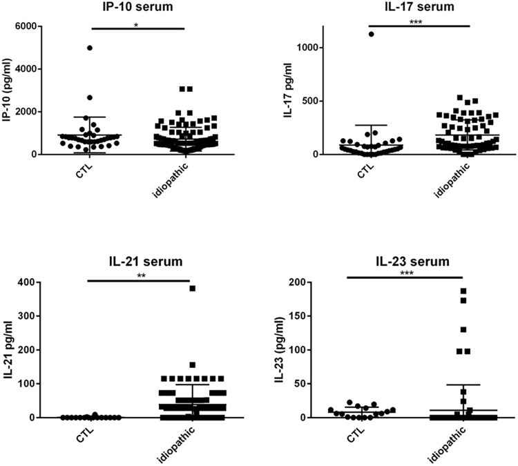
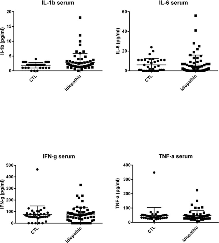
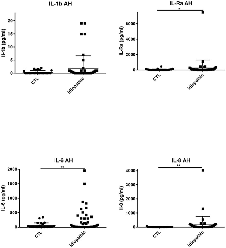
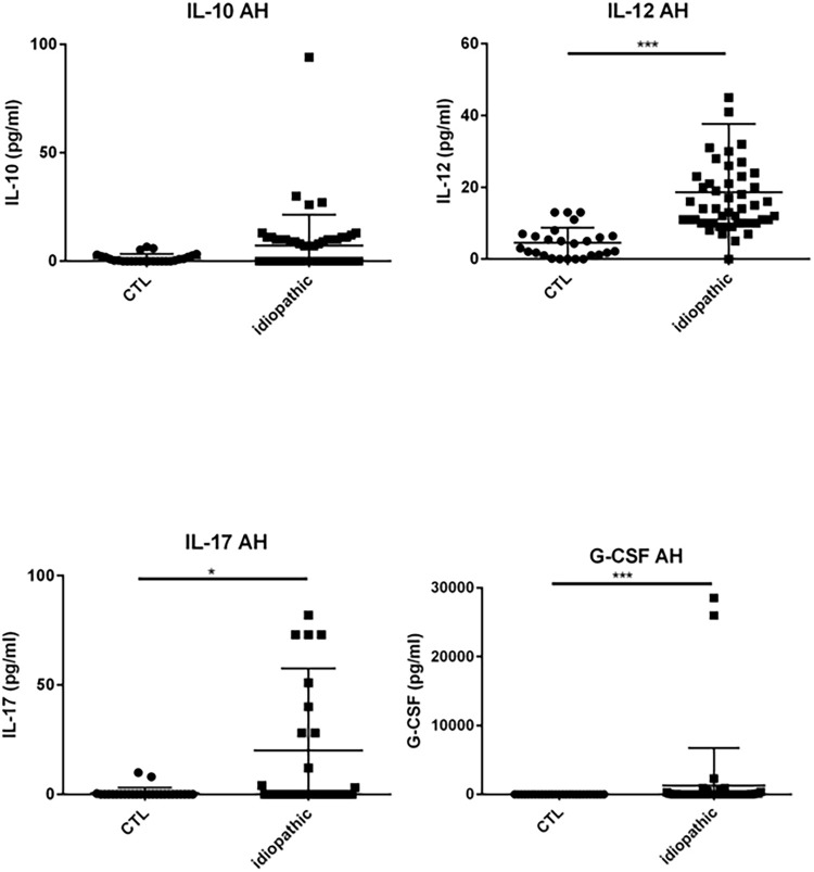
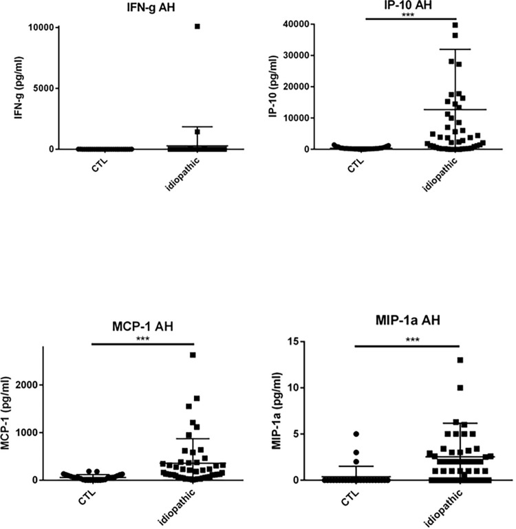
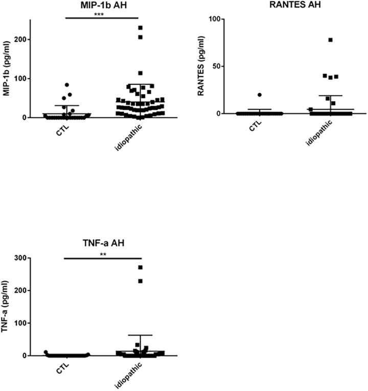
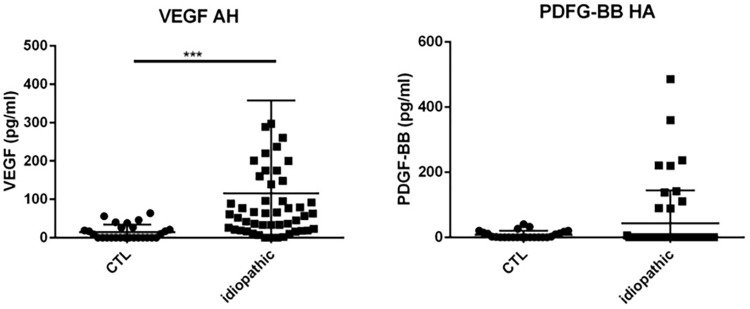
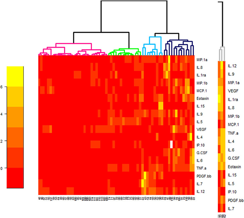
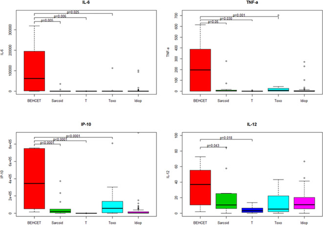
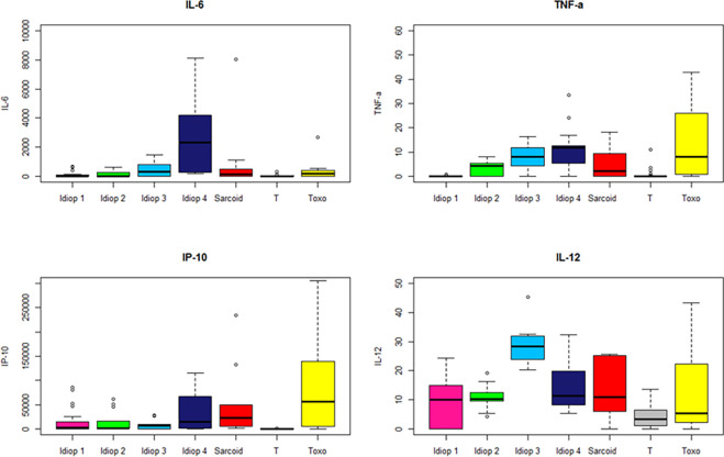
To investigate which cytokines, chemokines and growth factors are involved in the immunopathogenesis of idiopathic uveitis, and whether cytokine profiles are associated with. Serum and aqueous humor (AH) samples of 75 patients with idiopathic uveitis were analyzed by multiplex immunoassay. Infectious controls consisted of 16 patients with ocular toxoplasmosis all confirmed by intraocular fluid analyses. Noninfectious controls consisted of 7 patients with Behçet disease related uveitis and 15 patients with sarcoidosis related uveitis. The control group consisted of AH and serum samples from 47 noninflammatory control patients with age-related cataract. In each sample, 27 immune mediators ± IL-21 and IL-23 were measured. In idiopathic uveitis, 13 of the 29 mediators, including most proinflammatory and vascular mediators such as IL-6, IL-8, IL-12, G-CSF, GM-CSF, MCP-1, IP-10, TNF-α and VEGF, were significantly elevated in the aqueous humor when compared to all controls. Moreover, IL-17, IP-10, and IL-21, were significantly elevated in the serum when compared to all controls. We clustered 4 subgroups of idiopathic uveitis using a statistical analysis of hierarchical unsupervised classification, characterized by the order of magnitude of concentrations of intraocular cytokines. The pathogenesis of idiopathic uveitis is characterized by the presence of predominantly proinflammatory cytokines and chemokines and vascular endothelial growth factor with high expression levels as compared to other causes of uveitis. There are indications for obvious Th-1/ IL21-Th17 pathways but also IL9-Th9 and increased IFN-γ-inducing cytokine (IL12) and IFN-γ-inducible CXC chemokine (IP-10). The combined data suggest that immune mediator expression is different among idiopathic uveitis. This study suggests various clusters among the idiopathic uveitis group rather than one specific uveitis entity.


Conflict of interest statement

The authors have declared that no competing interests exist.

Figures

Fig 1
Fig 1. Dot plots of immune mediators, IL-17 and IP-10 in the serum.
IL-17 and IP-10 in the serum are increased in patients with idiopathic uveitis (n = 63) compared with serum of noninflammatory controls patients (cataract age-related) (CTL) (n = 47). Medians levels for IL-21 and IL-23 in the serum: elevated for IL-21 and decreased for IL-23 in patients with idiopathic uveitis (N = 68) as compared with noninflammatory controls patients (cataract age-related) (CTL) (N = 16). * = P<0.05; ** = P<0.01; *** = P<0.001.
Fig 2
Fig 2. Dot plots of immune mediators, IL-1β, IL-6, IFN-γ and TNF-α. IL-1β, IL-6, IFN-γ and TNF-α in serum of noninflammatory controls patients (cataract age-related) (CTL) (N = 36) as compared with immune mediators in the serum of patients with idiopathic uveitis (n = 51) but differences in both groups are not significant.
Note that few isolated patients with idiopathic uveitis had cytokines increased in serum samples. * = P<0.05; ** = P<0.01; *** = P<0.001.
Fig 3
Fig 3. Dot plots of immune mediators: IL-1β, IL1-Rα, IL-6, IL-8, in aqueous humor of patients with idiopathic uveitis (N = 64) as compared with immune mediators in the aqueous humor of noninflammatory controls patients (cataract age-related) (N = 36).
Note that few isolated patients with idiopathic uveitis had an immune mediator elevated. * = P<0.05; ** = P<0.01; *** = P<0.001.
Fig 4
Fig 4. Dot plots of immune mediators: IL-10, IL-12, IL-17, G-CSF, in aqueous humor of patients with idiopathic uveitis (N = 64) as compared with immune mediators in the aqueous humor of noninflammatory controls patients (cataract age-related) (N = 36).
Note that few isolated patients with idiopathic uveitis had an immune mediator elevated. * = P<0.05; ** = P<0.01; *** = P<0.001.
Fig 5
Fig 5. Dot plots of immune mediators: IFN-γ, IP-10, MCP-1, MIP-1α, in aqueous humor of patients with idiopathic uveitis (N = 64) as compared with immune mediators in the aqueous humor of noninflammatory controls patients (cataract age-related) (N = 36).
Note that few isolated patients with idiopathic uveitis had an immune mediator elevated. * = P<0.05; ** = P<0.01; *** = P<0.001.
Fig 6
Fig 6. Dot plots of immune mediators: MIP-1β, RANTES, TNF-α, in aqueous humor of patients with idiopathic uveitis (N = 64) as compared with immune mediators in the aqueous humor of noninflammatory controls patients (cataract age-related) (N = 36).
Note that few isolated patients with idiopathic uveitis had an immune mediator elevated. * = P<0.05; ** = P<0.01; *** = P<0.001.
Fig 7
Fig 7. Dot plots of immune mediators: VEGF and PDGF-BB, in aqueous humor of patients with idiopathic uveitis (N = 64) as compared with immune mediators in the aqueous humor of noninflammatory controls patients (cataract age-related) (N = 36).
Note that few isolated patients with idiopathic uveitis had an immune mediator elevated. * = P<0.05; ** = P<0.01; *** = P<0.001.
Fig 8
Fig 8. Heat map analysis comparing aqueous humor cytokine concentrations in 64 the idiopathic uveitis samples and 47 non-inflammatory controls (age-related cataracts).
Only significant different cytokines in idiopathic uveitis compared with controls were considered. Cytokine concentrations are depicted as colors ranging from red to orange to yellow, indicating low, intermediate, and high concentration, respectively, relative to the mean cytokine concentration. The phenotype of the samples is indicated by the label on the right as well as by the colored bar on the top. Samples are separated in one of four clusters (pink, green, blue and black) indicated on the top.
Fig 9
Fig 9. Boxplots of 4 immune mediators significantly elevated in the aqueous humor of patients with idiopathic uveitis (Idiop): IL-6, TNF-α, IL-12 and IP-10, as compared to immune mediators in the aqueous humor of patients with uveitis related to Behçet disease, sarcoidosis, TU (Toxo) and noninflammatory controls (T).
Significant P values are noted in the upper part of each graph. A P value < 0.05 was considered significant.
Fig 10
Fig 10. Boxplots of 4 immune mediators significantly elevated in the aqueous humor of patients within 4 sub-groups of idiopathic uveitis (Idiop 1, 2, 3 and 4): IL-6, TNF-α, IL-12 and IP-10, as compared to immune mediators in the aqueous humor of patients with uveitis related to sarcoidosis, TU (Toxo) and noninflammatory controls (T).
Significant P values are noted in the upper part of each graph. A P value < 0.05 was considered significant.

References

    1. Jaffe GJ, Dick AD, Brézin AP, Nguyen QD, Thorne JE, Kestelyn P, et al.. Adalimumab in Patients with Active Noninfectious Uveitis. N Engl J Med. 2016. Sep;375(10):932–43. doi: 10.1056/NEJMoa1509852 - DOI - PubMed
    1. Nguyen QD, Merrill PT, Jaffe GJ, Dick AD, Kurup SK, Sheppard J, et al.. Adalimumab for prevention of uveitic flare in patients with inactive non-infectious uveitis controlled by corticosteroids (VISUAL II): a multicentre, double-masked, randomised, placebo-controlled phase 3 trial. Lancet. 2016. Sep;388(10050):1183–92. doi: 10.1016/S0140-6736(16)31339-3 - DOI - PubMed
    1. Luger D, Caspi RR. New perspectives on effector mechanisms in uveitis. Semin Immunopathol. 2008. Apr;30(2):135–43. doi: 10.1007/s00281-008-0108-5 - DOI - PMC - PubMed
    1. Luger D, Caspi RR. New perspectives on effector mechanisms in uveitis. Semin Immunopathol. 2008. Apr;30(2):135–43. doi: 10.1007/s00281-008-0108-5 - DOI - PMC - PubMed
    1. Caspi RR. Ocular autoimmunity: the price of privilege? Immunol Rev. 2006. Oct;213:23–35. doi: 10.1111/j.1600-065X.2006.00439.x - DOI - PubMed

MeSH terms