. 2021 Dec 22;14(1):13.
doi: 10.3390/v14010013.

Adenovirus Co-Opts Neutrophilic Inflammation to Enhance Transduction of Epithelial Cells

Affiliations

Adenovirus Co-Opts Neutrophilic Inflammation to Enhance Transduction of Epithelial Cells

James M Readler et al. Viruses. .

Abstract

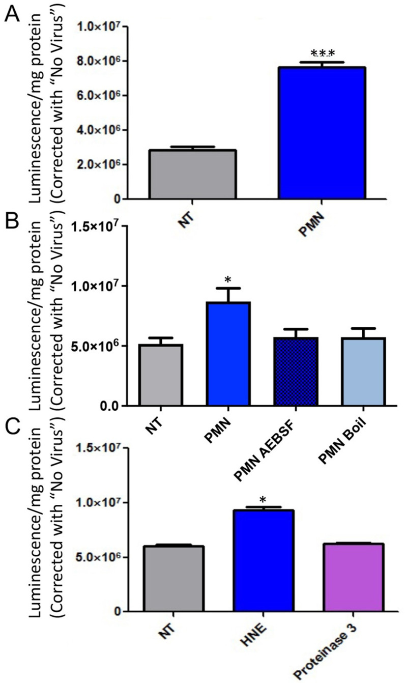
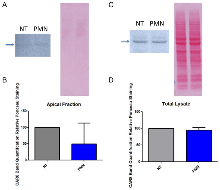
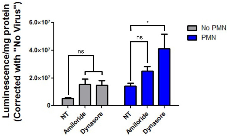
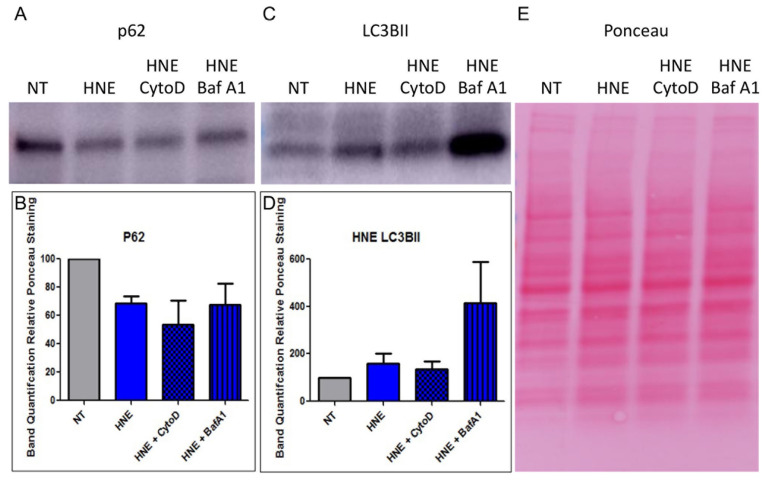
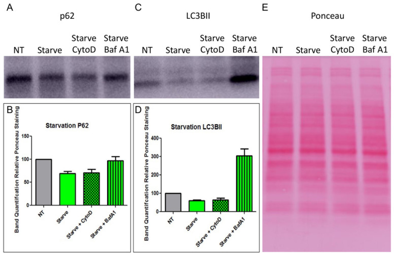
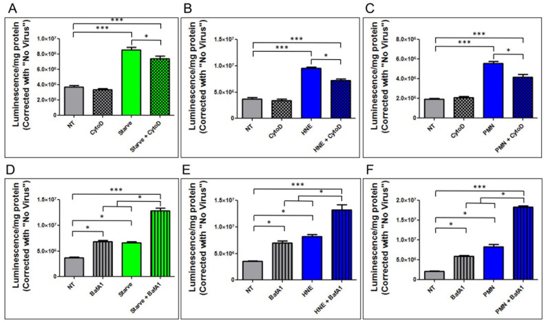
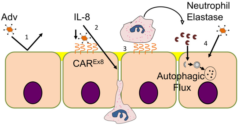
Human adenoviruses (HAdV) cause a variety of infections in human hosts, from self-limited upper respiratory tract infections in otherwise healthy people to fulminant pneumonia and death in immunocompromised patients. Many HAdV enter polarized epithelial cells by using the primary receptor, the Coxsackievirus and adenovirus receptor (CAR). Recently published data demonstrate that a potent neutrophil (PMN) chemoattractant, interleukin-8 (IL-8), stimulates airway epithelial cells to increase expression of the apical isoform of CAR (CAREx8), which results in increased epithelial HAdV type 5 (HAdV5) infection. However, the mechanism for PMN-enhanced epithelial HAdV5 transduction remains unclear. In this manuscript, the molecular mechanisms behind PMN mediated enhancement of epithelial HAdV5 transduction are characterized using an MDCK cell line that stably expresses human CAREx8 under a doxycycline inducible promoter (MDCK-CAREx8 cells). Contrary to our hypothesis, PMN exposure does not enhance HAdV5 entry by increasing CAREx8 expression nor through activation of non-specific epithelial endocytic pathways. Instead, PMN serine proteases are responsible for PMN-mediated enhancement of HAdV5 transduction in MDCK-CAREx8 cells. This is evidenced by reduced transduction upon inhibition of PMN serine proteases and increased transduction upon exposure to exogenous human neutrophil elastase (HNE). Furthermore, HNE exposure activates epithelial autophagic flux, which, even when triggered through other mechanisms, results in a similar enhancement of epithelial HAdV5 transduction. Inhibition of F-actin with cytochalasin D partially attenuates PMN mediated enhancement of HAdV transduction. Taken together, these findings suggest that HAdV5 can leverage innate immune responses to establish infections.

Keywords: MDCK epithelial cells; adenovirus; autophagy; entry; human neutrophil elastase; neutrophil.


Conflict of interest statement

The authors declare no conflict of interest. The funders had no role in the design of the study; in the collection, analyses, or interpretation of data; in the writing of the manuscript, or in the decision to publish the results.

Figures

Figure 1
Figure 1
Exposure to primary human neutrophils leads to increased AdV5 transduction, inhibition of neutrophil serine proteases ablates PMN mediated enhancement of epithelial AdV5 transduction, and addition of human neutrophil elastase alone enhances AdV5 transduction of MDCK-CAREx8 epithelia. (A) MDCK-CAREx8 epithelia were induced with doxycycline and 24 h later exposed to PMN. Cells were infected with AdV-LacZ, and beta galactosidase assay was performed the following day to measure AdV transduction. Data are presented as means ± standard errors (SEM) from quadruplicate samples in 13 independent experiments. *** p < 0.001 in two tailed Students t-test. (B) Freshly isolated PMN were treated with serine protease inhibitor AEBSF or vehicle control in HBSS at 37 °C or heat killed by placing in a boiling water bath for 30 min. Confluent monolayers of MDCK-CAREx8 epithelia in 24-well plates were then exposed to these PMN before infection with AdV5-LacZ and performing beta galactosidase assay the following day. (C) Confluent monolayers of MDCK-CAREx8 epithelia in 24-well plates were exposed to human neutrophil elastase, neutrophil proteinase 3, or vehicle control before infection with AdV5-LacZ and subsequent beta galactosidase assay. Error bars represent SEM of 3 independent experiments, each performed in quadruplicate. * p < 0.05 by one-way ANOVA and Bonferroni post-hoc test. NT, non-treated; all other treatments were compared to NT.
Figure 2
Figure 2
PMN exposure does not significantly change apical or total expression of CAREx8 in MDCK-CAREx8 epithelia. MDCK-CAREx8 cells were grown in 6-well transwell inserts until they were fully polarized and then exposed to PMN. Apical surface proteins were marked with sulfo-NHS-biotin and cell lysates were run through neutravidin beads to create an apical protein fraction. Cell lysates containing apical (A) and total (C) cell proteins were subjected SDS-PAGE, Ponceau staining, and subsequent Western blotting with antibody specific to CAREx8. Band quantifications are depicted for apical (B) and total (D) CAREx8 staining relative to Ponceau loading control. Error bars represent SEM from 3 independent experiments. NT, non-treated.
Figure 3
Figure 3
Inhibition of macropinocytosis and dynamin-dependent endocytosis pathways does not ablate PMN-mediated enhancement of epithelial AdV5 transduction. MDCK-CAREx8 cells were pretreated with amiloride or dynasore in serum-free media before exposure to PMN and subsequent infection with AdV5-LacZ, followed by beta-galactosidase activity assay the following day. Error bars represent SEM from 3 independent experiments each performed in triplicate. * p < 0.05 by one-way ANOVA and Bonferroni post-hoc test. NT, non-treated, ns, not significant.
Figure 4
Figure 4
HNE treatment results in activation of autophagic flux in MDCK-CAREx8 cells. MDCK-CAREx8 cells were pretreated with cytochalasin D or bafilomycin A1 and subsequently treated with HNE. The MDCK-CAREx8 cells were then washed thoroughly before performing Western blotting on the samples. Representative Western blot data of (A) anti-p62-treated, (C) anti-LC3BII-treated blots, as well as (E) Ponceau staining are shown. Band quantifications of (B) p62 bands relative Ponceau staining and (D) LC3BII bands relative to Ponceau staining are shown. Error bars represent SEM from 3 independent experiments each performed in quadruplicate. NT, non-treated.
Figure 5
Figure 5
HBSS starvation results in activation of autophagic flux in MDCK-CAREx8 cells. MDCK-CAREx8 cells were starved in HBSS+/+ for 4 h and then treated with cytochalasin D or bafilomycin A1. The MDCK-CAREx8 cells were then washed thoroughly before performing Western blotting on the samples. Representative Western blot data of (A) anti-p62 treated and (C) anti-LC3BII treated blots as well as (E) Ponceau staining are shown. Band quantifications of (B) p62 bands relative Ponceau staining and (D) LC3BII bands relative to Ponceau staining are shown. Error bars represent SEM from 3 independent experiments each performed in quadruplicate. NT, non-treated.
Figure 6
Figure 6
Cytochalasin D partially ablates and bafilomycin A1 further exacerbates enhancement of epithelial AdV5 transduction. MDCK-CAREx8 cells were either (A) exposed to HBSS starvation and then treated with cytochalasin D before treatment with (B) HNE or (C) PMN. MDCK-CAREx8 cells were then either (D) exposed to HBSS starvation and concurrently treated with bafilomycin A1 or treated with bafilomycin A1 and then subsequently treated with (E) HNE or (F) PMN. In all cases, the MDCK-CAREx8 cells were subsequently infected with AdV5-LacZ. Beta-galactosidase activity assay was performed the following day to measure AdV5 transduction. Error bars represent SEM from 3 independent experiments each performed in quadruplicate. * p < 0.05, *** p < 0.001 by one-way ANOVA and Bonferroni post-hoc test. NT, non-treated.
Figure 7
Figure 7
Summary model of how innate immune factors regulate epithelial susceptibility to AdV5 infection. (1) Under normal circumstances, polarized epithelia express very low levels of apical CAREx8, making them relatively resistant to apical AdV infection. (2) AdV-5 triggers release of proinflammatory cytokine IL-8, which induces apical expression of CAREx8 and (3) transepithelial migration of neutrophils. (4) Once neutrophils arrive, release of HNE further enhances AdV-5 transduction of epithelia through a process that involves activation of epithelial autophagic flux.

References

    1. Lynch J.P., 3rd, Kajon A.E. Adenovirus: Epidemiology, Global Spread of Novel Serotypes, and Advances in Treatment and Prevention. Semin. Respir. Crit. Care Med. 2016;37:586–602. - PMC - PubMed
    1. Lion T. Adenovirus infections in immunocompetent and immunocompromised patients. Clin. Microbiol. Rev. 2014;27:441–462. doi: 10.1128/CMR.00116-13. - DOI - PMC - PubMed
    1. Florescu D.F., Hoffman J.A., on behalf of the AST Infectious Diseases Community of Practice Adenovirus in solid organ transplantation. Am. J. Transpl. 2013;13((Suppl. S4)):206–211. doi: 10.1111/ajt.12112. - DOI - PubMed
    1. Florescu D.F., Schaenman J.M., on behalf of the AST Infectious Diseases Community of Practice Adenovirus in solid organ transplant recipients: Guidelines from the American Society of Transplantation Infectious Diseases Community of Practice. Clin. Transpl. 2019;33:e13527. doi: 10.1111/ctr.13527. - DOI - PubMed
    1. Vora S.B., Brothers A.W., Englund J.A. Renal Toxicity in Pediatric Patients Receiving Cidofovir for the Treatment of Adenovirus Infection. J. Pediatr. Infect. Dis. Soc. 2017;6:399–402. doi: 10.1093/jpids/pix011. - DOI - PubMed

Publication types

LinkOut - more resources