. 2022 Jan 5;14(1):88.
doi: 10.3390/v14010088.

Influenza A Virus Infection Activates NLRP3 Inflammasome through Trans-Golgi Network Dispersion

Affiliations

Influenza A Virus Infection Activates NLRP3 Inflammasome through Trans-Golgi Network Dispersion

Kannu Priya Pandey et al. Viruses. .

Abstract

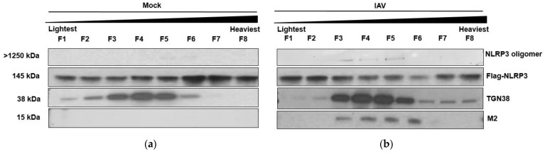
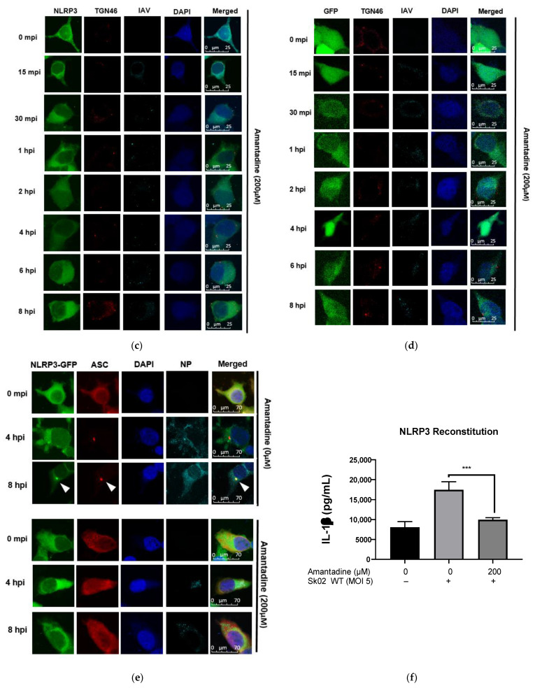
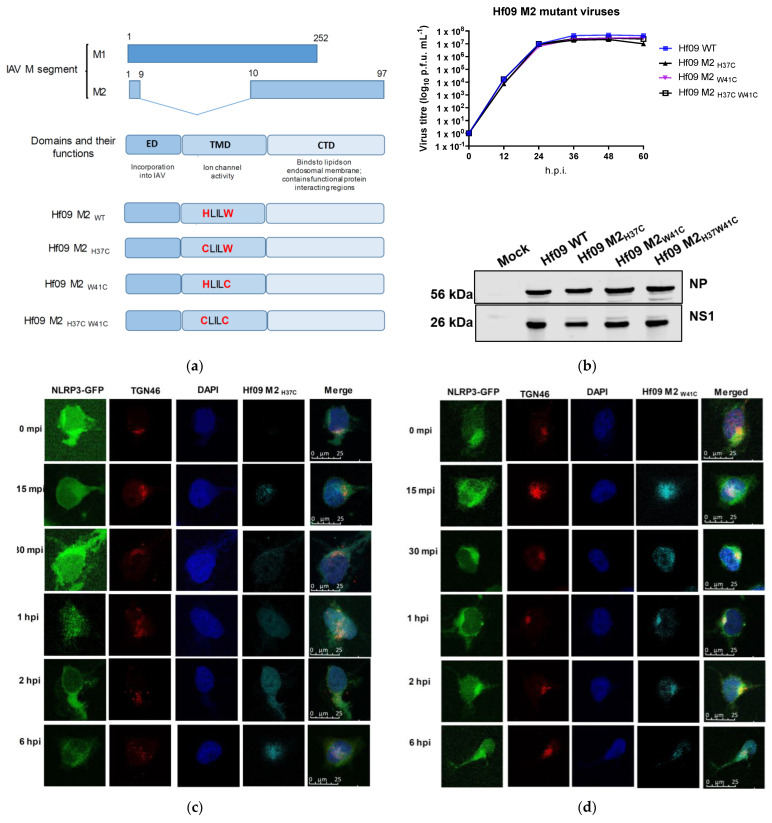
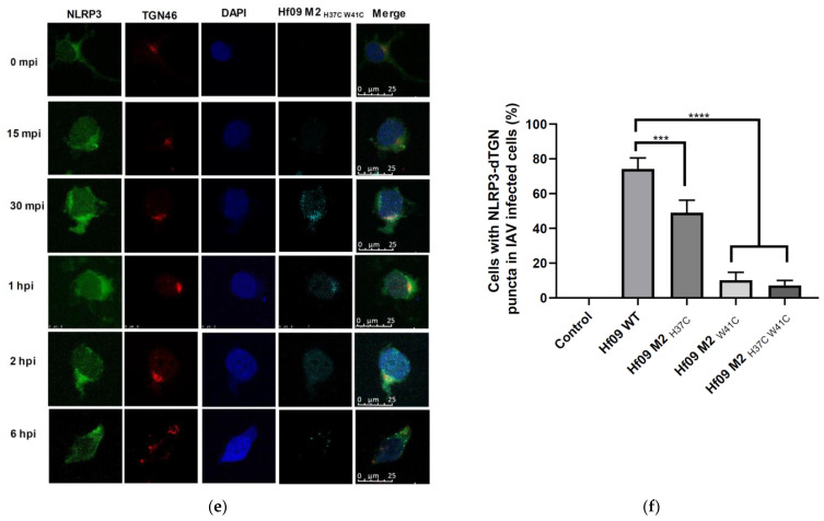
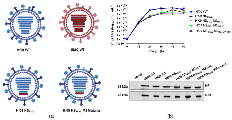
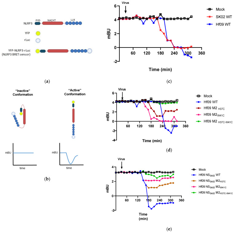
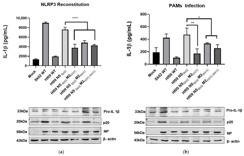
The NLRP3 inflammasome consists of NLRP3, ASC, and pro-caspase-1 and is an important arm of the innate immune response against influenza A virus (IAV) infection. Upon infection, the inflammasome is activated, resulting in the production of IL-1β and IL-18, which recruits other immune cells to the site of infection. It has been suggested that in the presence of stress molecules such as nigericin, the trans-Golgi network (TGN) disperses into small puncta-like structures where NLRP3 is recruited and activated. Here, we investigated whether IAV infection could lead to TGN dispersion, whether dispersed TGN (dTGN) is responsible for NLRP3 inflammasome activation, and which viral protein is involved in this process. We showed that the IAV causes dTGN formation, which serves as one of the mechanisms of NLRP3 inflammasome activation in response to IAV infection. Furthermore, we generated a series of mutant IAVs that carry mutations in the M2 protein. We demonstrated the M2 proton channel activity, specifically His37 and Trp41 are pivotal for the dispersion of TGN, NLRP3 conformational change, and IL-1β induction. The results revealed a novel mechanism behind the activation and regulation of the NLRP3 inflammasome in IAV infection.

Keywords: M2 ion channel protein; NLRP3 inflammasome; influenza A virus; trans-Golgi network.


Conflict of interest statement

The authors declare no conflict of interest. The funders had no role in the design of the study; in the collection, analyses, or interpretation of data; in the writing of the manuscript, or in the decision to publish the results.

Figures

Figure 1
Figure 1
IAV infection induces the formation of dispersed trans-Golgi network. (a) Schematic representation of Flag-NLRP3-GFP construct. (b) HEK293T cells were transfected with different combinations of plasmids expressing porcine NLRP3, ASC, pro-caspase-1, and pro-IL-1β as indicated. At 12 h.p.t., the cells were mock-infected or infected with Sk02 at an MOI of 10. Porcine IL-1β from the cell-free supernatants at 12 h.p.i was measured by ELISA (two-way ANOVA; **** p < 0.0001; ns- Not significant). (c) HeLa cells were co-transfected with ASC together with either GFP or NLRP3-GFP and were stimulated with LPS (200 ng/mL) for 80 min. The cells were fixed, permeabilized, blocked, and probed with appropriate antibodies, followed by DAPI staining. GFP/NLRP3-GFP (green), ASC (red), and nucleus (blue) were visualized by confocal microscopy. Scale bar, 50 μm. (d) HeLa cells expressing NLRP3-GFP were infected with Sk02 at an MOI of 10. At 80 m.p.i, the cells were subjected to immunofluorescence for NLRP3-GFP (green), TGN (red), IAV (cyan), and nucleus (blue). Scale bar, 50 μm.
Figure 1
Figure 1
IAV infection induces the formation of dispersed trans-Golgi network. (a) Schematic representation of Flag-NLRP3-GFP construct. (b) HEK293T cells were transfected with different combinations of plasmids expressing porcine NLRP3, ASC, pro-caspase-1, and pro-IL-1β as indicated. At 12 h.p.t., the cells were mock-infected or infected with Sk02 at an MOI of 10. Porcine IL-1β from the cell-free supernatants at 12 h.p.i was measured by ELISA (two-way ANOVA; **** p < 0.0001; ns- Not significant). (c) HeLa cells were co-transfected with ASC together with either GFP or NLRP3-GFP and were stimulated with LPS (200 ng/mL) for 80 min. The cells were fixed, permeabilized, blocked, and probed with appropriate antibodies, followed by DAPI staining. GFP/NLRP3-GFP (green), ASC (red), and nucleus (blue) were visualized by confocal microscopy. Scale bar, 50 μm. (d) HeLa cells expressing NLRP3-GFP were infected with Sk02 at an MOI of 10. At 80 m.p.i, the cells were subjected to immunofluorescence for NLRP3-GFP (green), TGN (red), IAV (cyan), and nucleus (blue). Scale bar, 50 μm.
Figure 2
Figure 2
ASC is recruited to the site of dTGN-NLRP3. HeLa cells expressing (a) NLRP3-GFP or (b) GFP were infected with Sk02 at an MOI of 10. At indicated time points post-infection, cells were subjected to immunofluorescence for NLRP3 (green), TGN46 (red), and IAV (cyan). Scale bar, 25 μm. HeLa cells expressing NLRP3-GFP (c) or GFP (d) were transfected with a plasmid expressing ASC-Myc for 24 h and then were infected with Sk02 at an MOI of 10. At indicated time points, the cells were subjected to immunofluorescence microscopy for NLRP3 (green), TGN46 (red), ASC (blue), and IAV (cyan). The white arrows indicate the co-localization of NLRP3 and ASC. Scale bar, 50 μm.
Figure 2
Figure 2
ASC is recruited to the site of dTGN-NLRP3. HeLa cells expressing (a) NLRP3-GFP or (b) GFP were infected with Sk02 at an MOI of 10. At indicated time points post-infection, cells were subjected to immunofluorescence for NLRP3 (green), TGN46 (red), and IAV (cyan). Scale bar, 25 μm. HeLa cells expressing NLRP3-GFP (c) or GFP (d) were transfected with a plasmid expressing ASC-Myc for 24 h and then were infected with Sk02 at an MOI of 10. At indicated time points, the cells were subjected to immunofluorescence microscopy for NLRP3 (green), TGN46 (red), ASC (blue), and IAV (cyan). The white arrows indicate the co-localization of NLRP3 and ASC. Scale bar, 50 μm.
Figure 3
Figure 3
M2 Protein influences dTGN formation before NLRP3 recruitment. HEK293T cells were transfected with Flag-NLRP3 for 48 h before being mock infected (a) or infected with Hf09 WT at MOI of 5 for 12 h (b). The cells were harvested and fractionated to isolate the light membrane fraction containing TGN. The fractions were then subjected to Western blot and probed for NLRP3, TGN, and M2. The cell fraction was also subjected to a native PAGE gel to test the presence of NLRP3 oligomers (top panels). HeLa cells expressing NLRP3-GFP (c) or GFP (d) were treated with Amantadine (200 μM) for 1 hr before being infected with Hf09 (MOI of 10). The cells were subjected to immunofluorescent microscopy for NLRP3 (green), TGN (red), IAV (cyan), and nucleus (blue). Scale bar, 25 μm (e) Cells expressing NLRP3-GFP were transfected with ASC-Myc for 12 h. The cells were then infected with Sk02 WT at an MOI of 10 in the presence or absence of Amantadine (200 μM). The cells were fixed at indicated time points and were subjected to immunofluorescent microscopy for NLRP3 (green), ASC (red), NP (cyan), and nucleus (blue). Scale bar, 70 μm; white arrows indicate ASC speck. (f) HEK293T was transfected with the various components of the NLRP3 inflammasome namely, NLRP3, ASC, pro-caspase-1, and pro-1L-1β, and incubated for 12 h. The media was then changed to basal DMEM containing 200 μM Amantadine and incubated for 1 hr before being infected with Sk02 WT (MOI of 5). The cell-free supernatant was harvested at 12 h.p.i. and the level of mature IL-1β released was measured by IL-1β ELISA (t-test, *** p = 0.0004).
Figure 3
Figure 3
M2 Protein influences dTGN formation before NLRP3 recruitment. HEK293T cells were transfected with Flag-NLRP3 for 48 h before being mock infected (a) or infected with Hf09 WT at MOI of 5 for 12 h (b). The cells were harvested and fractionated to isolate the light membrane fraction containing TGN. The fractions were then subjected to Western blot and probed for NLRP3, TGN, and M2. The cell fraction was also subjected to a native PAGE gel to test the presence of NLRP3 oligomers (top panels). HeLa cells expressing NLRP3-GFP (c) or GFP (d) were treated with Amantadine (200 μM) for 1 hr before being infected with Hf09 (MOI of 10). The cells were subjected to immunofluorescent microscopy for NLRP3 (green), TGN (red), IAV (cyan), and nucleus (blue). Scale bar, 25 μm (e) Cells expressing NLRP3-GFP were transfected with ASC-Myc for 12 h. The cells were then infected with Sk02 WT at an MOI of 10 in the presence or absence of Amantadine (200 μM). The cells were fixed at indicated time points and were subjected to immunofluorescent microscopy for NLRP3 (green), ASC (red), NP (cyan), and nucleus (blue). Scale bar, 70 μm; white arrows indicate ASC speck. (f) HEK293T was transfected with the various components of the NLRP3 inflammasome namely, NLRP3, ASC, pro-caspase-1, and pro-1L-1β, and incubated for 12 h. The media was then changed to basal DMEM containing 200 μM Amantadine and incubated for 1 hr before being infected with Sk02 WT (MOI of 5). The cell-free supernatant was harvested at 12 h.p.i. and the level of mature IL-1β released was measured by IL-1β ELISA (t-test, *** p = 0.0004).
Figure 4
Figure 4
IAV M2 ion channel protein activates NLRP3. (a) Schematic representation of M segment-encoded proteins, as well as the mutations made on M2 protein. (b) MDCK-M2 cells were infected with WT or one of the M2 defective Hf09 viruses at an MOI of 0.001. The supernatant was collected every 12 h and was subjected to plaque assays to measure the viral titers. Viral protein expression was determined in the infected cells harvested at 36 h.p.i. by Western blotting. Cells expressing NLRP3-GFP were infected with Hf09 M2H37C (c), Hf09 M2W41C (d), or Hf09 M2H37C-W41C (e) at an MOI of 10. At the indicated time points, the cells were fixed and subjected to immunofluorescent microscopy for NLRP3-GFP (green), TGN (red), IAV (cyan), and nucleus (blue). Scale bar, 25 μm. (f) The number of cells showing dTGN was counted from 100 IAV-infected cells (1 h.p.i.) in 5 different areas. (two-sided t-test, *** p = 0.0004; **** p < 0.0001).
Figure 4
Figure 4
IAV M2 ion channel protein activates NLRP3. (a) Schematic representation of M segment-encoded proteins, as well as the mutations made on M2 protein. (b) MDCK-M2 cells were infected with WT or one of the M2 defective Hf09 viruses at an MOI of 0.001. The supernatant was collected every 12 h and was subjected to plaque assays to measure the viral titers. Viral protein expression was determined in the infected cells harvested at 36 h.p.i. by Western blotting. Cells expressing NLRP3-GFP were infected with Hf09 M2H37C (c), Hf09 M2W41C (d), or Hf09 M2H37C-W41C (e) at an MOI of 10. At the indicated time points, the cells were fixed and subjected to immunofluorescent microscopy for NLRP3-GFP (green), TGN (red), IAV (cyan), and nucleus (blue). Scale bar, 25 μm. (f) The number of cells showing dTGN was counted from 100 IAV-infected cells (1 h.p.i.) in 5 different areas. (two-sided t-test, *** p = 0.0004; **** p < 0.0001).
Figure 5
Figure 5
Generation of Hf09 NSSk02 virus reassortants. (a) Schematic diagram of genome composition of WT and mutant viruses. Hf09 NSSK02 virus has the background of Hf09 WT (Blue) with NS segment replaced with that from Sk02 (Red) virus. Hf09 NSSk02 M2 mutant viruses (turquoise) are derivatives of Hf09 NSSK02 wherein the 7th segment contains various mutations. (b) MDCK- M2 cells were infected with Hf09 NSSk02 WT and the respective Hf09 NSSk02 M2 mutant viruses at an MOI of 0.001. The supernatant collected at different time points was subjected to plaque assay. Viral protein expression from infected cells (MOI of 1, at 36 h.p.i.) was determined by Western blotting (lower panel) (p.f.u.—plaque forming unit; time p.i.—Time post infection).
Figure 6
Figure 6
IAV induces NLRP3 conformational change upon infection. (a) Schematic representation illustrating the various components of the NLRP3 BRET sensor and (b) how the change in conformation of NLRP3 affects BRET signals (the two blue lines indicate BRET signals). HEK293T cells were transfected with pcDNA3.1- YFP-NLRP3-Luc for 24 h. The cells were then treated with Coelanterazine-h (5 μM). After 30 min, cells were infected with the following series of viruses at MOI of 1: Hf09 and Sk02 wild-type viruses (c) Hf09 virus and its derivatives containing M2 mutation (d), or Hf09 NSSk02 virus and its derivatives containing M2 mutation (e). The signal was recorded every 15 min and BRET signal is expressed as mBU (milli-BRET unit). The black arrow represents when the virus was added.
Figure 7
Figure 7
His37 and Trp41 in M2 affect IL-1β production. (a) HEK293T cells were transfected with different combinations of plasmids expressing porcine NLRP3, ASC, pro-caspase-1, and pro-IL-1β. At 12 h.p.t., the cells were mock-infected or infected with the viruses as indicated at an MOI of 5. Porcine IL-1β from the cell-free supernatants at 12 h.p.i. was measured by ELISA. The expression of pro-IL-1β, the active caspase-1, p20, NP, and β-actin was measured by Western blotting in the cell lysates (t-test, **** p < 0.0001). (b) PAMs were infected with various viruses as indicated at an MOI of 1 for 24 h. The cell-free supernatant was collected to measure the IL-1β level by ELISA and the cell pellet was lysed and checked for the level of pro-IL-1β, p20, NP, and β-actin by Western blotting (t-test, ** p = 0.0033; * p = 0.0339).
Figure 8
Figure 8
Schematic representation of the working model. Upon endocytosis of IAV, the pH of the endosome (pH 6) activates the IAV M2 ion channel protein leading to the release of vRNP into the cytoplasm. The vRNP then translocates into the nucleus to undergo transcription and translation. The newly translated M2 protein travels from the ER through the Golgi apparatus and reaches TGN. The pH of the TGN (pH 6) causes a pH disparity in the pH-sensitive organelle which leads to the dispersion of the TGN (dTGN). The NLRP3 molecules then travel to the newly formed dTGN where the NLRP3 undergoes conformational changes, oligomerization, and becomes active. The ASC molecule is then recruited to the site of dTGN-NLRP3, where the NLRP3 binds to ASC from its PYD domain. The NLRP3–ASC complex then leaves the dTGN and goes on to form the inflammasome, while the dTGN goes on to fuse with the cell membrane. The active NLRP3 inflammasome then leads to the production of active caspase-1 which then converts pro-IL-1β to mature IL-1β. The mature IL-1β is then released into the extracellular matrix, which then signals other immune cells to travel to the site of infection.

Similar articles

Cited by

References

    1. Mogensen T.H. Pathogen Recognition and Inflammatory Signaling in Innate Immune Defenses. Clin. Microbiol. Rev. 2009;22:240–273. doi: 10.1128/CMR.00046-08. - DOI - PMC - PubMed
    1. Amarante-Mendes G.P., Adjemian S., Branco L.M., Zanetti L., Weinlich R., Bortoluci K.R. Pattern Recognition Receptors and the Host Cell Death Molecular Machinery. Front. Immunol. 2018;9:2379. doi: 10.3389/fimmu.2018.02379. - DOI - PMC - PubMed
    1. Swanson K.V., Deng M., Ting J.P.-Y. The NLRP3 inflammasome: Molecular activation and regulation to therapeutics. Nat. Rev. Immunol. 2019;19:477–489. doi: 10.1038/s41577-019-0165-0. - DOI - PMC - PubMed
    1. Malik A., Kanneganti T.-D. Inflammasome activation and assembly at a glance. J. Cell Sci. 2017;130:3955–3963. doi: 10.1242/jcs.207365. - DOI - PMC - PubMed
    1. Kelley N., Jeltema D., Duan Y., He Y. The NLRP3 Inflammasome: An Overview of Mechanisms of Activation and Regulation. Int. J. Mol. Sci. 2019;20:3328. doi: 10.3390/ijms20133328. - DOI - PMC - PubMed

Publication types

MeSH terms

LinkOut - more resources