. 2022 Jan 9;14(1):113.
doi: 10.3390/v14010113.

SARS-like Coronaviruses in Horseshoe Bats (Rhinolophus spp.) in Russia, 2020

Affiliations

SARS-like Coronaviruses in Horseshoe Bats (Rhinolophus spp.) in Russia, 2020

Sergey Alkhovsky et al. Viruses. .

Abstract

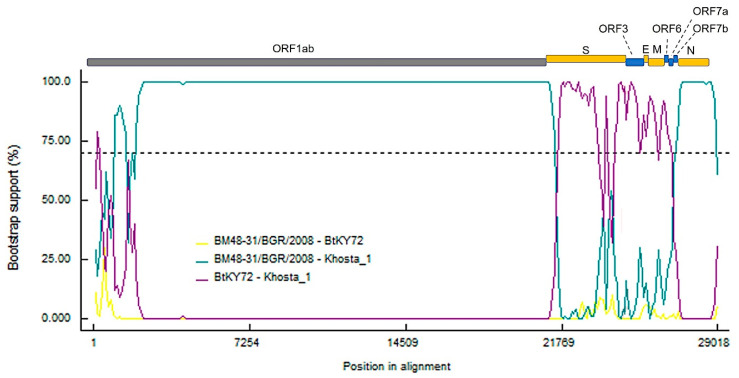
We found and genetically described two novel SARS-like coronaviruses in feces and oral swabs of the greater (R. ferrumequinum) and the lesser (R. hipposideros) horseshoe bats in southern regions of Russia. The viruses, named Khosta-1 and Khosta-2, together with related viruses from Bulgaria and Kenya, form a separate phylogenetic lineage. We found evidence of recombination events in the evolutionary history of Khosta-1, which involved the acquisition of the structural proteins S, E, and M, as well as the nonstructural genes ORF3, ORF6, ORF7a, and ORF7b, from a virus that is related to the Kenyan isolate BtKY72. The examination of bats by RT-PCR revealed that 62.5% of the greater horseshoe bats in one of the caves were positive for Khosta-1 virus, while its overall prevalence was 14%. The prevalence of Khosta-2 was 1.75%. Our results show that SARS-like coronaviruses circulate in horseshoe bats in the region, and we provide new data on their genetic diversity.

Keywords: Rhinolophus; SARS-CoV; SARS-CoV-2; SARS-CoV-like viruses; bat SARS-like coronaviruses; coronavirus; horseshoe bats; viral metagenomics; zoonotic viruses.


Conflict of interest statement

The authors declare no conflict of interest.

Figures

Figure 1
Figure 1
Map of the region where bat samples were collected. The location of Sochi National Park and the surrounding area is shown in gray.
Figure 2
Figure 2
Simplot analysis of Khosta-1 and Khosta-2 with SARS-CoV, SARS-CoV-2, and related viruses. RaTG13, HKU3, and Rs672 were used as representatives of bat SARS-CoV-like viruses from Asia. (A) Khosta-1 was used as a query sequence, and SARS-CoV, RaTG13, HKU3, Rs672, and SARS-CoV-2 were used as reference sequences. (B) Khosta-2 was used as a query sequence, and SARS-CoV, RaTG13, HKU3, Rs672, and SARS-CoV-2 were used as reference sequences. The sequences of the ORF8 gene, which is absent in Khosta-1 and Khosta-2, were removed from alignment before analysis. (C) Khosta-1 was used as a query sequence, and Khosta-2, BM48-31/BGR/2008, and BtKy72 viruses were used as reference sequences. (D) Khosta-2 was used as a query sequence, and Khosta-1, BM48-31/BGR/2008, and BtKY72 were used as reference sequences. The analysis was performed using the Kimura (two-parameter) model, with a window size of 1000 bases and a step size of 100 bases.
Figure 2
Figure 2
Simplot analysis of Khosta-1 and Khosta-2 with SARS-CoV, SARS-CoV-2, and related viruses. RaTG13, HKU3, and Rs672 were used as representatives of bat SARS-CoV-like viruses from Asia. (A) Khosta-1 was used as a query sequence, and SARS-CoV, RaTG13, HKU3, Rs672, and SARS-CoV-2 were used as reference sequences. (B) Khosta-2 was used as a query sequence, and SARS-CoV, RaTG13, HKU3, Rs672, and SARS-CoV-2 were used as reference sequences. The sequences of the ORF8 gene, which is absent in Khosta-1 and Khosta-2, were removed from alignment before analysis. (C) Khosta-1 was used as a query sequence, and Khosta-2, BM48-31/BGR/2008, and BtKy72 viruses were used as reference sequences. (D) Khosta-2 was used as a query sequence, and Khosta-1, BM48-31/BGR/2008, and BtKY72 were used as reference sequences. The analysis was performed using the Kimura (two-parameter) model, with a window size of 1000 bases and a step size of 100 bases.
Figure 3
Figure 3
Phylogenetic trees inferred using maximum likelihood method according to an analysis of nucleotide sequences of the RdRp gene (2766 nt) (A), nucleotide sequences of the S gene (3822 nt (SARS-CoV-2 numbering)) (B), and nucleotide sequences of N gene (1257 nt) (C) of certain sarbecoviruses. The percentage of trees in which the associated taxa clustered together is shown next to the branches (values higher 50% are shown). SARS-CoV and SARS-CoV-2 are marked by black circles; Khosta-1 and Khosta-2, described in the present work, are marked by red circles. The trees were inferred using GTR + G + I model with 1000 bootstrap replicates using the MEGAX.
Figure 3
Figure 3
Phylogenetic trees inferred using maximum likelihood method according to an analysis of nucleotide sequences of the RdRp gene (2766 nt) (A), nucleotide sequences of the S gene (3822 nt (SARS-CoV-2 numbering)) (B), and nucleotide sequences of N gene (1257 nt) (C) of certain sarbecoviruses. The percentage of trees in which the associated taxa clustered together is shown next to the branches (values higher 50% are shown). SARS-CoV and SARS-CoV-2 are marked by black circles; Khosta-1 and Khosta-2, described in the present work, are marked by red circles. The trees were inferred using GTR + G + I model with 1000 bootstrap replicates using the MEGAX.
Figure 4
Figure 4
Results of bootscan analysis of recombination events in Khosta-1 genome using RDP5 software. The analysis was performed using the Jukes and Cantor (1969) model, with a window size of 1000 bases, a step size of 100 bases and a number of bootstrep replicates of 100 as implemented in RDP5 program. Cutoff percentage (70%) shown by dashed line. The colors of the lines correspond to different pairs of analyzed viruses as indicated in the legend.
Figure 5
Figure 5
Amino acid alignment of the receptor-binding motif (RBM) of the receptor-binding domain (RBD) of the S protein of Khosta-1 and Khosta-2 of certain sarbecoviruses. Five positions (442, 472, 479, 480, and 487; SARS-CoV Urbani numbering) in the RBM that are thought to be important for adaptation of SARS-CoV-like viruses to the human ACE2 receptor [17] are shown in bold. Bat SARS-CoV-like viruses that are capable or incapable of utilizing the ACE2 receptor are marked with “ACE2(+)” or “ACE2(−)”, respectively (A). Amino acid alignment of the region around the S1/S2 cleavage site in SARS-CoV-2, SARS-CoV, Khosta-1, Khosta-2, and certain bat SARS-CoV-like viruses. Four-amino acids insertion (PRRA) that forms polybasic furin cleavage site in SARS-CoV-2 is highlighted with a red border (B).

References

    1. Lau S.K., Woo P.C., Li K.S., Huang Y., Tsoi H.W., Wong B.H., Wong S.S., Leung S.Y., Chan K.H., Yuen K.Y. Severe acute respiratory syndrome coronavirus-like virus in Chinese horseshoe bats. Proc. Natl. Acad. Sci. USA. 2005;102:14040–14045. doi: 10.1073/pnas.0506735102. - DOI - PMC - PubMed
    1. Li W., Shi Z., Yu M., Ren W., Smith C., Epstein J.H., Wang H., Crameri G., Hu Z., Zhang H., et al. Bats are natural reservoirs of SARS-like coronaviruses. Science. 2005;310:676–679. doi: 10.1126/science.1118391. - DOI - PubMed
    1. De Groot R.J., Baker S.C., Baric R., Enjuanes L., Gorbalenya A.E., Holmes K.V., Perlman S., Poon L., Rottier P.J.M., Talbot P.J., et al. Family Coronaviridae. In: King A.M., Adams M.J., Carstens E.B., Lefkowitz E.J., editors. Virus Taxonomy: Classification and Nomenclature of Viruses: Ninth Report of the International Committee on Taxonomy of Viruses. Elsevier; London, UK: 2012. pp. 806–828.
    1. Fan Y., Zhao K., Shi Z.-L., Zhou P. Bat Coronaviruses in China. Viruses. 2019;11:210. doi: 10.3390/v11030210. - DOI - PMC - PubMed
    1. Rihtarič D., Hostnik P., Steyer A., Grom J., Toplak I. Identification of SARS-like coronaviruses in horseshoe bats (Rhinolophus hipposideros) in Slovenia. Arch. Virol. 2010;155:507–514. doi: 10.1007/s00705-010-0612-5. - DOI - PMC - PubMed

Publication types

Substances

LinkOut - more resources