. 2022 Jan 14;14(1):153.
doi: 10.3390/v14010153.

Actin Contributes to the Hyperexpression of Baculovirus Polyhedrin (polh) and p10 as a Component of Transcription Initiation Complex (TIC)

Affiliations

Actin Contributes to the Hyperexpression of Baculovirus Polyhedrin (polh) and p10 as a Component of Transcription Initiation Complex (TIC)

Nan Chen et al. Viruses. .

Abstract

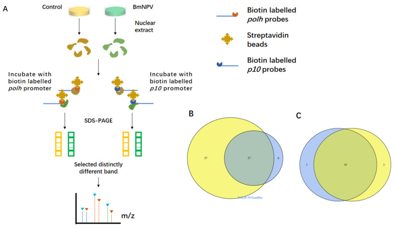
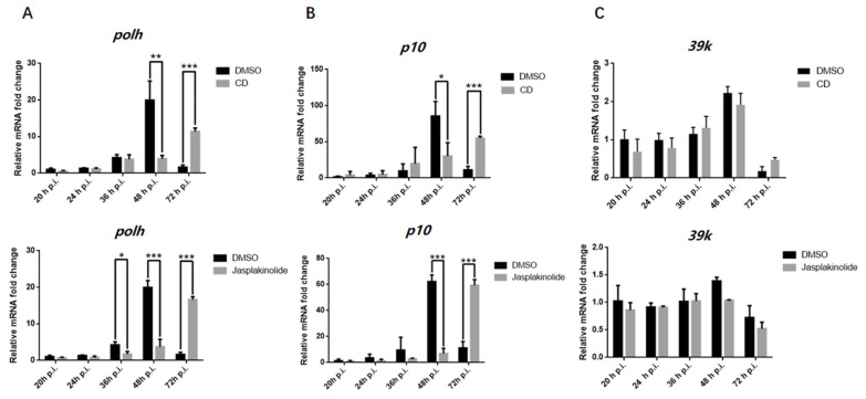
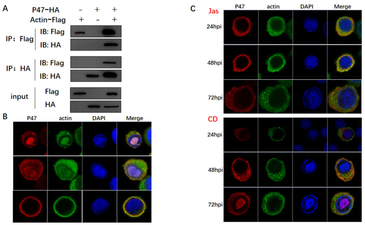
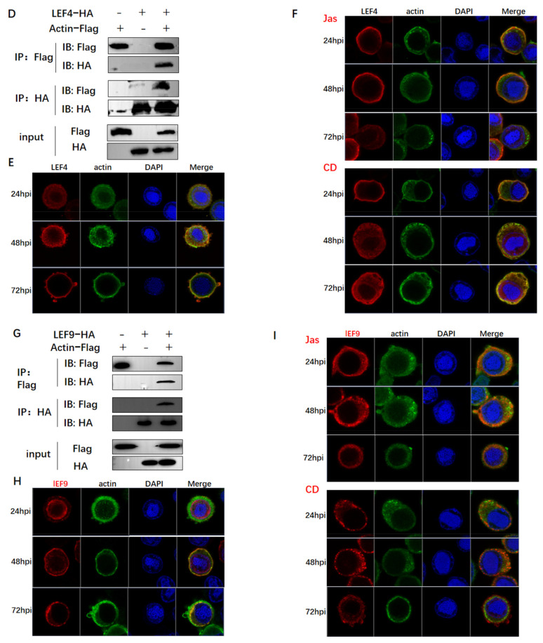
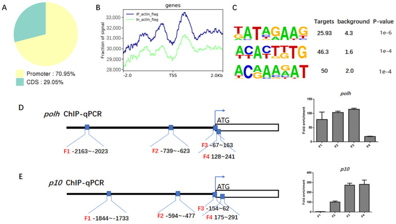
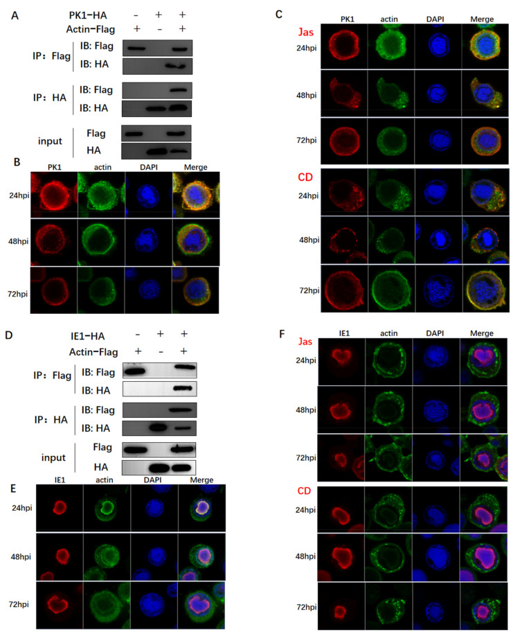
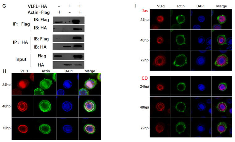
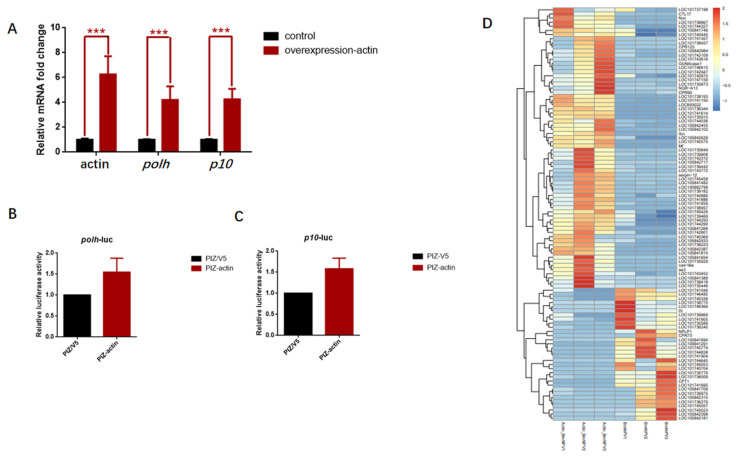
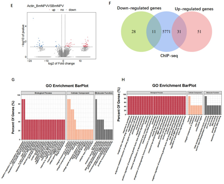
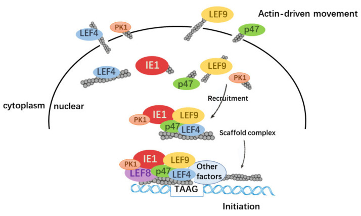
Hyperexpression of polh and p10, two very late genes, is one of the remarkable characteristics in the baculovirus life cycle. However, the mechanisms underlying the hyperexpression of these two genes are still incompletely understood. In this study, actin was identified as a highly potential binding partner of polh and p10 promoters by conducting DNA pull-down and LC-MS/MS analyses. Inhibiting actin dynamics delayed and decreased the transcription of polh and p10. Actin interacted with viral RNA polymerase and transcription regulators, and the nuclear import of viral polymerase was inhibited with the disruption of actin dynamics. Simultaneously, the high enrichment of actin in polh and p10 promoters discovered via a chromatin immunoprecipitation (ChIP) assay indicated that actin was a component of the viral polymerase TIC. Moreover, overexpression of actin surprisingly upregulated the expression of luciferase (Luc) under the control of polh and p10 promoters. Taken together, actin participated in the hyperexpression of polh and p10 as a component of TIC. These results facilitate the promotion of the expression efficiency of foreign genes in the baculovirus expression vector system (BEVS).

Keywords: BEVS; actin; baculovirus; hyperexpression.


Conflict of interest statement

The authors declare no conflict of interest.

Figures

Figure 1
Figure 1
MS analysis of the binding proteins to the polh and p10 promoter. (A) Schematic of our approach. (B) Venn diagram displaying the summary statistics of the host proteins bound to polh promoter and p10 promoter. Total proteins identified in polh are indicated by a blue circle and proteins identified in p10 are indicated by a yellow circle. (C) As in (B), summary statistics of the viral proteins bound to polh promoter and p10 promoter.
Figure 2
Figure 2
RT-qPCR analysis of the effect on polh and p10 transcription caused by CD and Jas. (A) Quantitative real-time PCR analysis of polh in BmN cells treated with CD, Jas, or DMSO (Ctrl) (n = 3; *, p < 0.05; **, p < 0.01; ***, p < 0.001). (B) Quantitative real-time PCR analysis of p10 in BmN cells treated with CD, Jas, or DMSO (Ctrl) (n = 3; *, p < 0.05; ***, p < 0.001). (C) Quantitative real-time PCR analysis of 39k in BmN cells treated with CD, Jas, or DMSO (Ctrl) (n = 3). The white columns are for the dimethyl sulfoxide (DMSO) treatment as control, the black columns are for CD and Jas treatment. The data indicate the means plus standard errors from three independent assays.
Figure 3
Figure 3
The analysis of the relationship between actin and viral polymerase. (A,D,G) Co-IP assays used to confirm interactions of actin with P47, LEF4, and LEF9. BmN cells coinfected with recombinant viruses vBm-actin-Flag and vBm-P47-HA, vBm-LEF4-HA, and vBm-LEF9-HA were lysed at 72 h p.i. and the proteins were immunoprecipitated with anti-Flag or anti-HA monoclonal antibody. The precipitates (Co-IP) were detected by Western blots with anti-Flag and anti-HA monoclonal antibody. The cell lysates (lysate input) were also examined by Western blots with an anti-HA rabbit polyclonal antibody or an anti-Flag rabbit monoclonal antibody. (B,E,H) Co-localization analysis of actin with P47, LEF4, and LEF9 by confocal microscopy. At the designated time points, BmN cells coinfected with recombinant viruses vBm-actin-Flag and vBm-P47-HA, vBm-LEF4-HA, and vBm-LEF9-HA were fixed, permeabilized, blocked, and incubated with rabbit anti-Flag antibody and mouse anti-HA antibody, followed by treatment with Alexa Fluor 546–conjugated goat anti-rabbit IgG and Alexa Fluor 488–conjugated goat anti-mouse IgG. (C,F,I) As in (B,E,H), at 15 h p.i., cells were treated with CD or Jas and then subjected to confocal microscopy.
Figure 3
Figure 3
The analysis of the relationship between actin and viral polymerase. (A,D,G) Co-IP assays used to confirm interactions of actin with P47, LEF4, and LEF9. BmN cells coinfected with recombinant viruses vBm-actin-Flag and vBm-P47-HA, vBm-LEF4-HA, and vBm-LEF9-HA were lysed at 72 h p.i. and the proteins were immunoprecipitated with anti-Flag or anti-HA monoclonal antibody. The precipitates (Co-IP) were detected by Western blots with anti-Flag and anti-HA monoclonal antibody. The cell lysates (lysate input) were also examined by Western blots with an anti-HA rabbit polyclonal antibody or an anti-Flag rabbit monoclonal antibody. (B,E,H) Co-localization analysis of actin with P47, LEF4, and LEF9 by confocal microscopy. At the designated time points, BmN cells coinfected with recombinant viruses vBm-actin-Flag and vBm-P47-HA, vBm-LEF4-HA, and vBm-LEF9-HA were fixed, permeabilized, blocked, and incubated with rabbit anti-Flag antibody and mouse anti-HA antibody, followed by treatment with Alexa Fluor 546–conjugated goat anti-rabbit IgG and Alexa Fluor 488–conjugated goat anti-mouse IgG. (C,F,I) As in (B,E,H), at 15 h p.i., cells were treated with CD or Jas and then subjected to confocal microscopy.
Figure 4
Figure 4
ChIP-seq and ChIP-qPCR analysis. (A) Statistics of distribution of actin-binding sites in the virus genome. (B) Distribution of actin-binding sites in the genic regions of the recombinant virus Bm-actin-Flag. (C) The top four most highly enriched motifs of actin in viral gene. Information regarding the most significant motifs of viral genes identified in the actin-binding peaks with Multiple EM for Motif Elicitation (MEME). (D,E) Verification of the ChIP-Seq results by ChIP-qPCR including four regions upstream of polh and p10 TSS. The primer pairs used in the RT-qPCR assay are represented with black lines (black boxes from left to right are F1 to F4). Enrichment of target DNA was represented as a percentage of input DNA. The values in each column are the means of three independent replicates and error bars represent the SEM.
Figure 5
Figure 5
The analysis of the relationship between actin and transcriptional regulators. (A,D,G) Co-IP assays used to confirm interactions of actin with PK1 and IE1. The method was the same as in Figure 3. (B,E,H) Co-localization analysis of actin with PK1 and IE1 by confocal microscopy. The method was the same as in Figure 3. (C,F,I) As in (B,E,H), at 15 h p.i., cells were treated with Jas or CD and then subjected to confocal microscopy.
Figure 5
Figure 5
The analysis of the relationship between actin and transcriptional regulators. (A,D,G) Co-IP assays used to confirm interactions of actin with PK1 and IE1. The method was the same as in Figure 3. (B,E,H) Co-localization analysis of actin with PK1 and IE1 by confocal microscopy. The method was the same as in Figure 3. (C,F,I) As in (B,E,H), at 15 h p.i., cells were treated with Jas or CD and then subjected to confocal microscopy.
Figure 6
Figure 6
Overexpression of actin stimulates polh and p10 transcription. (A) Quantitative real-time PCR analysis of actin, polh, and p10 in BmN cells overexpressing actin or empty vector (Ctrl) (n = 3, ***, p < 0.001). (B,C) The relative luciferase expression activity under the control of polh and p10. (D) Heatmap showing the fold change for differentially expressed host genes in cells overexpressing actin and control cells expressing empty vector. (E) Scatter plot of global mRNA expression in cells overexpressing actin and control by polyA(+) RNA-seq. Number of up- and downregulated mRNAs is shown. (F) Venn diagram representing overlap between differentially expressed genes (up and down) and the enrichment host genes of actin from ChIP-seq. (G,H) The functional annotations of the overlapped genes in (E).
Figure 6
Figure 6
Overexpression of actin stimulates polh and p10 transcription. (A) Quantitative real-time PCR analysis of actin, polh, and p10 in BmN cells overexpressing actin or empty vector (Ctrl) (n = 3, ***, p < 0.001). (B,C) The relative luciferase expression activity under the control of polh and p10. (D) Heatmap showing the fold change for differentially expressed host genes in cells overexpressing actin and control cells expressing empty vector. (E) Scatter plot of global mRNA expression in cells overexpressing actin and control by polyA(+) RNA-seq. Number of up- and downregulated mRNAs is shown. (F) Venn diagram representing overlap between differentially expressed genes (up and down) and the enrichment host genes of actin from ChIP-seq. (G,H) The functional annotations of the overlapped genes in (E).
Figure 7
Figure 7
Summary model for the regulatory roles of actin play in baculovirus polymerase transcription. A model of how actin dynamics assists in the hyperexpression in the baculovirus expression vector system.

Similar articles

References

    1. Kost T.A., Condreay J.P., Jarvis D.L. Baculovirus as versatile vectors for protein expression in insect and mammalian cells. Nat. Biotechnol. 2005;23:567–575. doi: 10.1038/nbt1095. - DOI - PMC - PubMed
    1. Vlak J.M., Klinkenberg A.F., Zaal K.J.M., Usmany M., Klinge-Roode E.C., Geervliet J.B.F., Roosien J., Van Lent J.W.M., Klinkenberg F. Functional studies on the p10 gene of Autographa californica nuclear polyhedrosis virus using a recombinant expressing a p10-beta-galactosidase fusion gene. Pt 4J. Gen. Virol. 1988;69:765–776. doi: 10.1099/0022-1317-69-4-765. - DOI - PubMed
    1. Blissard G.W., Rohrmann G.F. Baculovirus diversity and molecular biology. Annu. Rev. Entomol. 1990;35:127–155. doi: 10.1146/annurev.en.35.010190.001015. - DOI - PubMed
    1. Felberbaum R.S. The baculovirus expression vector system: A commercial manufacturing platform for viral vaccines and gene therapy vectors. Biotechnol. J. 2015;10:702–714. doi: 10.1002/biot.201400438. - DOI - PMC - PubMed
    1. van Oers M.M., Pijlman G.P., Vlak J.M. Thirty years of baculovirus-insect cell protein expression: From dark horse to mainstream technology. Pt 1J. Gen. Virol. 2015;96:6–23. doi: 10.1099/vir.0.067108-0. - DOI - PubMed

Publication types

LinkOut - more resources