

New Trends in Melanoma Detection Using Neural Networks: A Systematic Review
- PMID: 35062458
- PMCID: PMC8778535
- DOI: 10.3390/s22020496
New Trends in Melanoma Detection Using Neural Networks: A Systematic Review
Abstract
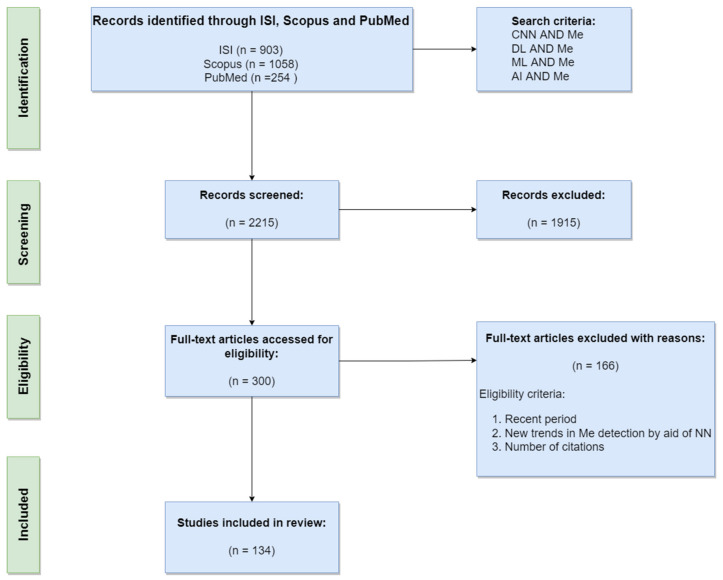
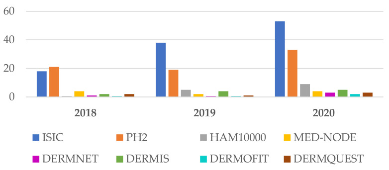
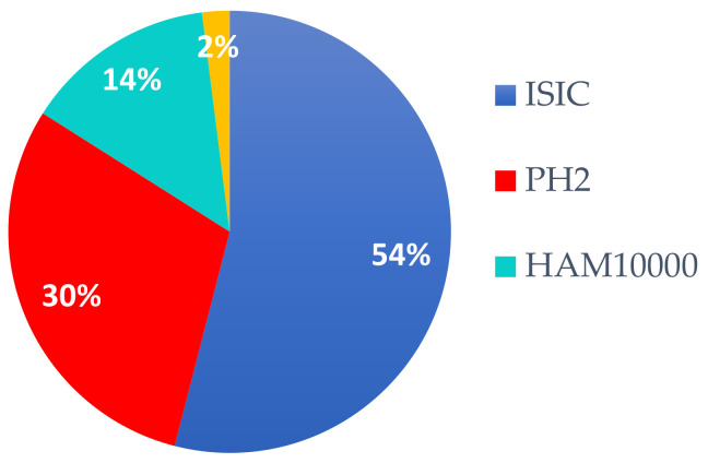
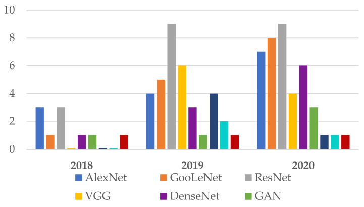
Due to its increasing incidence, skin cancer, and especially melanoma, is a serious health disease today. The high mortality rate associated with melanoma makes it necessary to detect the early stages to be treated urgently and properly. This is the reason why many researchers in this domain wanted to obtain accurate computer-aided diagnosis systems to assist in the early detection and diagnosis of such diseases. The paper presents a systematic review of recent advances in an area of increased interest for cancer prediction, with a focus on a comparative perspective of melanoma detection using artificial intelligence, especially neural network-based systems. Such structures can be considered intelligent support systems for dermatologists. Theoretical and applied contributions were investigated in the new development trends of multiple neural network architecture, based on decision fusion. The most representative articles covering the area of melanoma detection based on neural networks, published in journals and impact conferences, were investigated between 2015 and 2021, focusing on the interval 2018-2021 as new trends. Additionally presented are the main databases and trends in their use in teaching neural networks to detect melanomas. Finally, a research agenda was highlighted to advance the field towards the new trends.
Keywords: deep learning; image classifiers; image processing; image segmentation; machine learning; melanoma detection; neural networks; review; skin lesion; statistic performances.
Conflict of interest statement
The authors declare no conflict of interest.
Figures
Similar articles
-
Automatic melanoma detection using an optimized five-stream convolutional neural network.Sci Rep. 2025 Jul 1;15(1):22404. doi: 10.1038/s41598-025-05675-w. Sci Rep. 2025. PMID: 40594676 Free PMC article.
-
Multiclass skin lesion classification and localziation from dermoscopic images using a novel network-level fused deep architecture and explainable artificial intelligence.BMC Med Inform Decis Mak. 2025 Jul 1;25(1):215. doi: 10.1186/s12911-025-03051-2. BMC Med Inform Decis Mak. 2025. PMID: 40597254 Free PMC article.
-
Integrating Patient Data Into Skin Cancer Classification Using Convolutional Neural Networks: Systematic Review.J Med Internet Res. 2021 Jul 2;23(7):e20708. doi: 10.2196/20708. J Med Internet Res. 2021. PMID: 34255646 Free PMC article.
-
Systematic review of approaches to detection and classification of skin cancer using artificial intelligence: Development and prospects.Comput Biol Med. 2024 Aug;178:108742. doi: 10.1016/j.compbiomed.2024.108742. Epub 2024 Jun 14. Comput Biol Med. 2024. PMID: 38875908
-
Melanoma Breslow Thickness Classification Using Ensemble-Based Knowledge Distillation With Semi-Supervised Convolutional Neural Networks.IEEE J Biomed Health Inform. 2025 Jan;29(1):443-455. doi: 10.1109/JBHI.2024.3465929. Epub 2025 Jan 7. IEEE J Biomed Health Inform. 2025. PMID: 39302772
Cited by
-
Refining skin lesions classification performance using geometric features of superpixels.Sci Rep. 2023 Jul 15;13(1):11463. doi: 10.1038/s41598-023-38706-5. Sci Rep. 2023. PMID: 37454166 Free PMC article.
-
Artificial Intelligence on Diagnostic Aid of Leprosy: A Systematic Literature Review.J Clin Med. 2023 Dec 28;13(1):180. doi: 10.3390/jcm13010180. J Clin Med. 2023. PMID: 38202187 Free PMC article. Review.
-
Neural Networks for the Detection of COVID-19 and Other Diseases: Prospects and Challenges.Bioengineering (Basel). 2023 Jul 18;10(7):850. doi: 10.3390/bioengineering10070850. Bioengineering (Basel). 2023. PMID: 37508877 Free PMC article.
-
circTADA2A inhibited SLC38A1 expression and suppresses melanoma progression through the prevention of CNBP trans-activation.PLoS One. 2024 Apr 18;19(4):e0301356. doi: 10.1371/journal.pone.0301356. eCollection 2024. PLoS One. 2024. PMID: 38635778 Free PMC article.
-
Skin Lesion Classification Using Collective Intelligence of Multiple Neural Networks.Sensors (Basel). 2022 Jun 10;22(12):4399. doi: 10.3390/s22124399. Sensors (Basel). 2022. PMID: 35746180 Free PMC article.
References
-
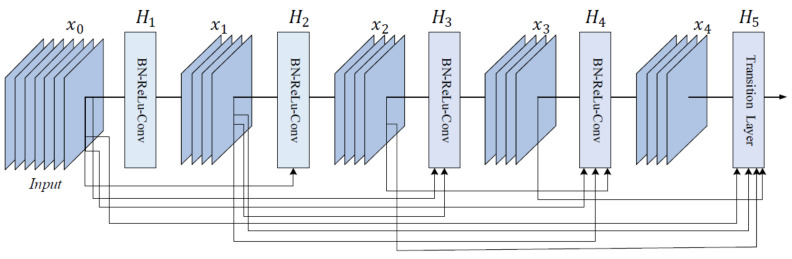
- Adegun A., Viriri S. FCN-based DenseNet framework for automated detection and classification of skin lesions in dermoscopy images. IEEE Access. 2020;8:150377–150396. doi: 10.1109/ACCESS.2020.3016651. - DOI
-
- Matthews N.H., Li W.Q., Qureshi A.A., Weinstock M.A., Cho E. Epidemiology of melanoma. In: Ward W.H., Farma J.M., editors. Cutaneous Melanoma: Etiology and Therapy [Internet] Codon Publications; Brisbane, Australia: 2017. - PubMed
Publication types
MeSH terms
LinkOut - more resources
Full Text Sources
Medical
