. 2022 Jan 3;10(1):71.
doi: 10.3390/vaccines10010071.

Elucidating the 3D Structure of a Surface Membrane Antigen from Trypanosoma cruzi as a Serodiagnostic Biomarker of Chagas Disease

Affiliations

Elucidating the 3D Structure of a Surface Membrane Antigen from Trypanosoma cruzi as a Serodiagnostic Biomarker of Chagas Disease

Flavio Di Pisa et al. Vaccines (Basel). .

Abstract

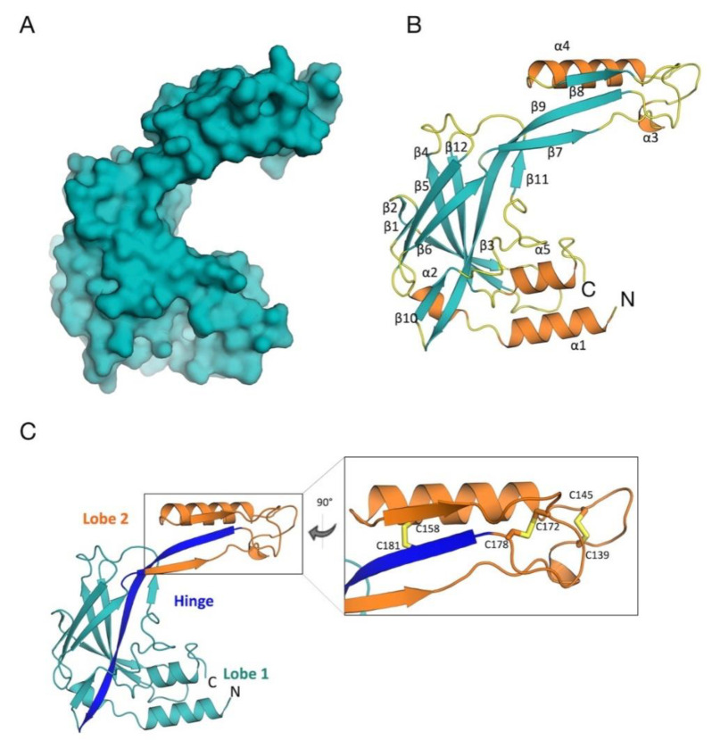
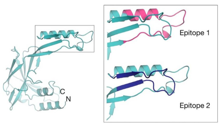
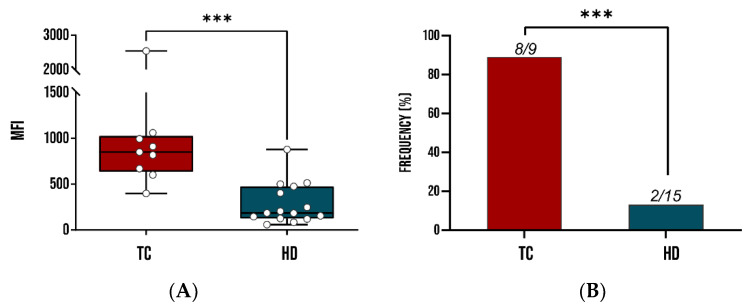
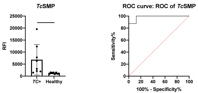
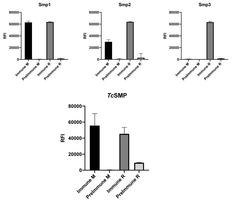
Chagas disease (CD) is a vector-borne parasitosis, caused by the protozoan parasite Trypanosoma cruzi, that affects millions of people worldwide. Although endemic in South America, CD is emerging throughout the world due to climate change and increased immigratory flux of infected people to non-endemic regions. Containing of the diffusion of CD is challenged by the asymptomatic nature of the disease in early infection stages and by the lack of a rapid and effective diagnostic test. With the aim of designing new serodiagnostic molecules to be implemented in a microarray-based diagnostic set-up for early screening of CD, herein, we report the recombinant production of the extracellular domain of a surface membrane antigen from T. cruzi (TcSMP) and confirm its ability to detect plasma antibodies from infected patients. Moreover, we describe its high-resolution (1.62 Å) crystal structure, to which in silico epitope predictions were applied in order to locate the most immunoreactive regions of TcSMP in order to guide the design of epitopes that may be used as an alternative to the full-length antigen for CD diagnosis. Two putative, linear epitopes, belonging to the same immunogenic region, were synthesized as free peptides, and their immunological properties were tested in vitro. Although both peptides were shown to adopt a structural conformation that allowed their recognition by polyclonal antibodies raised against the recombinant protein, they were not serodiagnostic for T. cruzi infections. Nevertheless, they represent good starting points for further iterative structure-based (re)design cycles.

Keywords: Chagas disease; Trypanosoma cruzi; immunodiagnostics; in silico epitope mapping; neglected tropical disease; peptide microarray; structural vaccinology.


Conflict of interest statement

The authors declare no conflict of interest. The funders had no role in the design of the study; in the collection, analyses, or interpretation of data; in the writing of the manuscript; or in the decision to publish the results.

Figures

Figure 1
Figure 1
3D structure of TcSMP. (A) Surface representation, illustrating the “C-shaped” organization of TcSMP. (B) Cartoon, secondary structure representation of TcSMP (β-strands in teal, α-helices in orange, 3-, 4-, and 5-turns and loops in yellow). The N and C-termini are indicated. (C) Stereo view of the TcSMP crystal structure. TcSMP is structurally organized into two lobes (colored in teal and orange), connected by a hinge portion, constituted by a β-strand and a connecting loop, depicted in blue. A detailed view of the region of lobe 2 that hosts the three disulfide bonds (sticks) is shown. This figure was generated using CCP4mg [54].
Figure 2
Figure 2
Structure-based TcSMP epitope mapping. The location of computationally predicted epitopes are mapped on the crystal structure of TcSMP. A zoom view of epitope 1 (residues 130–156; pink) and epitope 2 (residues 163–182; blue) is shown. This figure was generated using CCP4mg [54].
Figure 3
Figure 3
Immunoreactivity studies of recombinant TcSMP in sera from patients infected by T. cruzi in comparison with healthy donors (HDs). A whisker plot comparing mean fluorescence intensity of mean fluorescence intensity (MFI) of TcSMP tested against the sera of 9 T. cruzi-infected patients and 15 HDs. (A) Each dot represents the MFI of a single patient. (B) Recognition frequency of TcSMP as determined by DELFIA assay. ***: statistical significance p < 0.0001 Student’s t-test and Fisher’s exact test, p < 0.0001 for MFIs and recognition frequencies. TC: T. cruzi patient sera; HD: healthy donors.
Figure 4
Figure 4
Immune sera reactivity potential of recombinant TcSMP. Left panel: unpaired t-test results for the detection of TcSMP-specific human IgG. Protein arrays were probed with sera (N = 8) from patients with confirmed T. cruzi infections (TC+) and healthy control patients (N = 14). Significative: p < 0.05.
Figure 5
Figure 5
Immune sera reactivity of predicted epitopes. Upper panels: ability of biotinylated peptides Smp1, Smp2, and Smp3 to recognize antibodies present in mouse immune sera (M) and rabbit (R) immune sera, following immunization with recombinant TcSMP. Lower panel: immune sera reactivity of mouse immune sera (M) and rabbit (R) immune sera, against the recombinant TcSMP protein.

References

    1. Schofield C.J., Jannin J., Salvatella R. The future of Chagas disease control. Trends Parasitol. 2006;22:583–588. doi: 10.1016/j.pt.2006.09.011. - DOI - PubMed
    1. WHO Chagas Disease (American Trypanosomiasis) [(accessed on 11 May 2021)]. Available online: https://www.who.int/news-room/fact-sheets/detail/chagas-disease-(america...
    1. CDC American Trypanosomiasis. [(accessed on 16 February 2021)]; Available online: https://www.cdc.gov/parasites/chagas/
    1. Lidani K.C.F., Andrade F.A., Bavia L., Damasceno F.S., Beltrame M.H., Messias-Reason I.J., Sandri T.L. Chagas Disease: From Discovery to a Worldwide Health Problem. Front. Public Health. 2019;7:166. doi: 10.3389/fpubh.2019.00166. - DOI - PMC - PubMed
    1. Stuart K., Brun R., Croft S., Fairlamb A., Gürtler R.E., McKerrow J., Reed S., Tarleton R. Kinetoplastids: Related protozoan pathogens, different diseases. J. Clin. Investig. 2008;118:1301–1310. doi: 10.1172/JCI33945. - DOI - PMC - PubMed

LinkOut - more resources